| PMC full text: | Published online 2012 Aug 13. doi: 10.1186/1742-2094-9-196
|
Figure 2

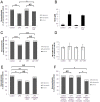
Disruption of CRT/LRP phagocytic signalling inhibits primary phagocytosis induced by LPS or Aβ. (A, C +
+ E) Co-cultures of cerebellar neurons and glia were treated with 100
 ng/ml LPS for 72
 hours in the presence of 1
 μg/ml CRT-blocking antibody (A), 250 nM RAP (C) or 1
 μg/ml LRP-blocking antibody (E). In (A) and (E) normal mouse IgG (mIgG) was added to control for non-specific effects of CRT- and LRP-blocking antibodies. In (C) and (I) ‘control’ refers to addition of PBS alone as RAP was dissolved in PBS prior to addition. Neuronal survival was quantified using Hoechst/PI staining after 72
 hours. (B, D
 +
+ F) Production of nitrite as a measure of microglial activation was measured in cell culture supernatants from experiments shown in (A) (B), (C) (D) and (E) (F). (G-J) Cerebellar co-cultures were treated with 250 nM Aβ peptide for 72
 hours in the presence of CRT-blocking antibody (G
 +
+ H) or RAP (I
 +
+ J) prior to quantification of neuronal survival (G
 +
+ I) and microglial density (H
 +
+ J). All data represent the mean value of three separate experiments (two replicates per experiment). Error bars represent SEM. Aβ, amyloid-β peptide1-42; CRT, calreticulin; LPS, lipopolysaccharide; LRP, low-density lipoprotein receptor-related protein; RAP, receptor-associated protein.


