Abstract
Unlabelled
Background
Mice from the MRL or "superhealing" strain have enhanced repair after acute injury to the skin, cornea, and heart. We now tested an admixture of the MRL genome and found that it altered the course of muscle pathology and cardiac function in a chronic disease model of skeletal and cardiac muscle. Mice lacking γ-sarcoglycan (Sgcg), a dystrophin-associated protein, develop muscular dystrophy and cardiomyopathy similar to their human counterparts with limb girdle muscular dystrophy. With disruption of the dystrophin complex, the muscle plasma membrane becomes leaky and muscles develop increased fibrosis.Methods
MRL/MpJ mice were bred with Sgcg mice, and cardiac function was measured. Muscles were assessed for fibrosis and membrane leak using measurements of hydroxyproline and Evans blue dye. Quantitative trait locus mapping was conducted using single nucleotide polymorphisms distinct between the two parental strains.Results
Introduction of the MRL genome reduced fibrosis but did not alter membrane leak in skeletal muscle of the Sgcg model. The MRL genome was also associated with improved cardiac function with reversal of depressed fractional shortening and the left ventricular ejection fraction. We conducted a genome-wide analysis of genetic modifiers and found that a region on chromosome 2 was associated with cardiac, diaphragm muscle and abdominal muscle fibrosis.Conclusions
These data are consistent with a model where the MRL genome acts in a dominant manner to suppress fibrosis in this chronic disease setting of heart and muscle disease.Free full text

The superhealing MRL background improves muscular dystrophy
Associated Data
Abstract
Background
Mice from the MRL or “superhealing” strain have enhanced repair after acute injury to the skin, cornea, and heart. We now tested an admixture of the MRL genome and found that it altered the course of muscle pathology and cardiac function in a chronic disease model of skeletal and cardiac muscle. Mice lacking γ-sarcoglycan (Sgcg), a dystrophin-associated protein, develop muscular dystrophy and cardiomyopathy similar to their human counterparts with limb girdle muscular dystrophy. With disruption of the dystrophin complex, the muscle plasma membrane becomes leaky and muscles develop increased fibrosis.
Methods
MRL/MpJ mice were bred with Sgcg mice, and cardiac function was measured. Muscles were assessed for fibrosis and membrane leak using measurements of hydroxyproline and Evans blue dye. Quantitative trait locus mapping was conducted using single nucleotide polymorphisms distinct between the two parental strains.
Results
Introduction of the MRL genome reduced fibrosis but did not alter membrane leak in skeletal muscle of the Sgcg model. The MRL genome was also associated with improved cardiac function with reversal of depressed fractional shortening and the left ventricular ejection fraction. We conducted a genome-wide analysis of genetic modifiers and found that a region on chromosome 2 was associated with cardiac, diaphragm muscle and abdominal muscle fibrosis.
Conclusions
These data are consistent with a model where the MRL genome acts in a dominant manner to suppress fibrosis in this chronic disease setting of heart and muscle disease.
Background
Murphy Roths Large (MRL) mice are an inbred mouse strain noted to have enhanced healing ability. This MRL strain was initially discovered because of its rapid ability to heal ear holes [1,2]. The MRL strain’s capacity to rapidly recover from injury has been seen for both digit wounding and corneal scarring [3,4]. The MRL strain has been reported to reduce scar formation after acute cardiac injury, including freeze injury and coronary artery ligation [5,6]. However, other studies have suggested that larger scale acute cardiac injury cannot be overcome by the MRL strain’s healing capacity [7-11]. In those injury settings where the MRL background induces more rapid healing, multiple mechanisms have been implicated to explain this phenomenon including decreased scar formation, altered inflammatory response, reduced apoptosis, increased proliferation, improved remodeling and, in some settings, enhanced stem cell function [4,6,12-15]. Genetic data support that many different mechanisms account for enhanced healing since more than 40 different genetic loci have been associated with aspects of the healing phenotype [14,16].
The dystrophin complex is composed of membrane-associated proteins that mediate membrane stability in heart and skeletal muscle. Mutations that disrupt expression of dystrophin or its associated proteins the sarcoglycans proteins cause progressive cardiac and skeletal muscle degeneration in humans and mouse models. At the cellular level, the loss of dystrophin or the sarcoglycans leads to a disrupted sarcolemmal membrane that is abnormally leaky. Membrane permeability results in increased intracellular calcium that triggers proteolysis and necrosis. An inflammatory response also contributes to muscle degeneration [17]. Muscle contraction is thought to provoke submicroscopic disruption of the sarcolemma, and in the absence of a normal dystrophin complex, this leads to dysfunction and destruction of cardiomyocytes and skeletal myofibers. The reduction in contractile cells and the presence of fibrotic scar tissue within the heart led to reduced cardiac contractile function and congestive heart failure.
Mouse models of dystrophin or sarcoglycan mutations recapitulate the basic pathological defects seen in human forms of these genetic disorders. Mice lacking γ-sarcoglycan were engineered by removing the first coding exon of the Sgcg gene and model muscular dystrophy [18]. We previously introduced the Sgcg allele into the DBA/2J genetic background and found that this background confers a more severe phenotype [19]. The enhanced severity is seen as increased scar formation, measured as hydroxyproline (HOP) content because this modified amino acid is a marker of collagen. In skeletal muscle, increased Evans blue dye uptake is monitored to reflect membrane leakiness [20]. Using these assays, we showed that the DBA/2J genetic background worsened the disease process [19].
Because of its role in wound healing and the basic similarities between common forms of cellular injury and what is seen in muscular dystrophy and cardiomyopathy, we introduced the MRL genome into the Sgcg model. We hypothesized that the indolent pace of cellular damage in this disorder could be abated by the MRL background. Sgcg mice were bred to MRL mice to generate Sgcg mice on a mixed genetic background with 50% contribution of the MRL strain. This breeding strategy produced Sgcg mice with 50% genetic background from the MRL strain and 50% background of the DBA/2J strain, and these mice are referred to SgcgMRL/D2. We found that a 50% contribution of the MRL genome reduced fibrosis in the heart and skeletal muscle, consistent with dominant genetic loci in the MRL background. Interestingly, the MRL genome did not consistently reduce membrane leak, suggesting that the MRL background does not exert its effect on myocyte membrane stability and instead acts downstream on remodeling. SgcgMRL/D2 mice had improved cardiac function so that they were now similar to wild-type mice. We conducted a genome-wide scan using informative polymorphisms and identified a region on chromosome 2 that was associated with fibrosis in cardiac, diaphragm and abdominal muscles. These data demonstrate that genes within the MRL background are modifiers for cardiopulmonary involvement in muscular dystrophy.
Methods
Animals
The Sgcg mouse was previously bred for ten generations into the DBA/2J strain (000671, Jackson Laboratory, Bar Harbor, ME) [18,19]. MRL/MpJ (000486, Jackson Laboratory, Bar Harbor, ME) mice were bred to the Sgcg animals to generate Sgcg+/− F1 mice on a 50% MRL/50% DBA/2J background. F1 mice were interbred to generate an F2 cohort. To increase the number of mutant animals, Sgcg mice from the F2 generation were bred again to produce additional Sgcg mice from an F3 generation. All mice were housed in uniform conditions in a single pathogen-free barrier facility. All animals used in this study were housed and treated in accordance with the standards set by the University of Chicago Animal Care and Use Committee. The number of mice used for histopathological analysis is shown in Table Table11.
Table 1
Number of mice analyzed
| Trait | Genotypes | ||
|---|---|---|---|
| Fibrosis | SgcgD2 | SgcgMRL/D2 | SgcgMRL/D2, 32wk |
| Quadriceps | 25 | 124 | 18 |
| Diaphragm | 25 | 45 | 10 |
| Heart | 27 | 125 | 9 |
| Membrane leak | SgcgD2 | SgcgMRL/D2 | SgcgMRL/D2, 32wk |
| Quadriceps | 37 | 149 | 15 |
| Gluteus | 11 | 76 | 15 |
| Triceps | 14 | 110 | 15 |
| Abdominals | 14 | 74 | 15 |
| Gastrocnemius/soleus | 14 | 111 | 15 |
| Central nuclei | SgcgD2 | SgcgMRL/D2 | |
| Quadriceps | 6 | 7 | |
| Diaphragm | 6 | 4 | |
| Fiber size variability | SgcgD2 | SgcgMRL/D2 | |
| Quadriceps | 7 | 9 | |
| Diaphragm | 6 | 4 | |
Mice were sacrificed at either 8 or 32 weeks for analysis. Muscles were used for either the Evans blue dye assay or the HOP assay. Dye uptake assays were performed on triceps muscles, gastrocnemius/soleus muscles and gluteus/hamstrings groups of muscles. Dye uptake was performed on half of each quadriceps muscle and half of the abdominal muscles since these muscles are of sufficient size to perform both dye uptake and fibrosis assays. Hydroxyproline (HOP) assays were performed on the diaphragm muscle, the cardiac ventricles isolated as a single unit, half of the abdominal muscles and half of each quadriceps muscle. The amount of muscle assayed varied with the size of the muscle and ranged from 30 mg for the diaphragm muscle to 300 mg for the gluteus/hamstring muscle group that included the semimembranosus, semitendinosus and biceps femoris.
Evans blue dye uptake assay for membrane leak
Evans blue dye (Sigma, E-2129) was performed as described [19,21]. Evans blue dye (Sigma, E-2129) was dissolved in phosphate-buffered saline at 10 mg/ml and injected intraperitoneally at 5 μl/g body weight. Twenty to 40 h later, the tissues were harvested, finely minced, weighed and incubated at 55°C in 1 ml formamide for 2 h before spectrophotometric absorbance was measured at 620 nm [22,23]. Results are reported as absorbance/mg tissue.
Hydroxyproline assay for fibrosis
The hydroxyproline (HOP) assay was performed as described [19,21,24]. The tissue was minced, weighed and hydrolyzed overnight in 2 ml of 6 M hydrochloric acid at 110°C. Ten μl of this hydrolysate was mixed with 150 μl isopropanol, then 75 μl of 1.4% chloramine-T (Sigma, St Louis, MO) in citrate buffer and oxidized at room temperature for 10 min. One ml of a 3:13 solution of Ehrlich’s reagent (3 g of 4-(dimethylamino) benzaldehyde, Sigma, St Louis, MO; 10 ml ethanol; 675 μl sulfuric acid) to isopropanol was added, mixed and incubated for 30 min at 55°C followed by extinction measurement at 558 nm. A standard curve (0–5000 nM, trans-4-hydroxy-L-proline, Sigma, St Louis, MO) was included in each assay. Results are reported as nM HOP/mg tissue.
Immunofluorescence microscopy
Tissues were flash frozen in liquid nitrogen-cooled isopentane and stored at 80°C; 7-μm sections were cut on a cryostat and fixed to slides in ice-cold 100% methanol. The following antibodies were used: dystrophin NCL-DYS2 (Novocastra/Leica), PH3 04–817 (Millipore), CD3 MON1003-1 (Monsanto), caspase 3 (BD Biosciences), MAC1 BD557395 (BD Biosciences) and eMHC F1.652 (ATCC). The TUNEL kit was from Millipore. Central nuclei and fiber size variability was determined blinded to genotype by analyzing ten randomly chosen fields of 40× magnification. Fiber size variability was compared using each animal’s coefficient of variability (standard deviation/mean).
Echocardiography
Twelve-week animals were evaluated by echocardiography as described [25,26]. Investigators were blinded to genotype. To avoid the stress associated with conscious restraint, anesthetized animals were studied. Anesthesia was induced by 1% isoflurane in a closed chamber (Ohmeda Fluotec 3; Matrix Medical, Orchard Park, NY) in 20% O2 delivered through a nose cone. Chest hairs were removed with a topical depilatory agent. Limb leads were attached for electrocardiogram gating, and the animals were imaged in the left lateral decubitus position with a Visual Sonics Vevo 770 machine using a 30-MHz high-frequency transducer. Body temperature was maintained using a heated imaging platform and warming lamps. Anesthesia was variably delivered to maintain heart rates throughout the procedure at a constant 380–420 beats per minute. Two-dimensional images were recorded in parasternal long- and short-axis projections, with guided M-mode recordings at the midventricular level in both views. LV cavity size and wall thickness were measured in at least three beats from each projection and averaged. LV wall thickness, interventricular septum (IVS) and posterior wall (PW) thickness, and internal dimensions at diastole and systole (LVIDd and LVIDs, respectively) were measured. LV fractional shortening [(LVIDd – LVIDs)/LVIDd] and relative wall thickness [(IVS thickness + PW thickness)/LVIDd] were calculated from the M-mode measurements.
Genetic and statistical analysis
Single nucleotide polymorphisms (SNPs, n = 1,701) informative between the parental DBA/2J and MRL/MpJ strains were genotyped in 80 SgcgMRL/D2F2-F4 Sgcg animals on the Illumina GoldenGate platform using the Mutation Mapping and Developmental Analysis Panel (MMDAP) [27] and the Mouse Universal Genotyping Array (GeneSeek, Neogen Corp., Lansing, MI) [28]. R package QTLRel was used to perform whole-genome quantitative trait locus (QTL) mapping for each of the membrane permeability and fibrosis phenotypes and using sex as a covariate [29,30]. Significance thresholds were determined by 1,000 permutation tests. The 1.5-LOD drop support interval was calculated using QTLRel. Tests of normality and other statistics were calculated in Prism (GraphPad). Build 37.1 was used for genomic analysis.
For HOP and dye uptake assays, data were analyzed by one-way, unpaired ANOVA with parametric methods followed by the Tukey multiple comparison post-test (Prism, Graphpad), and p < 0.05 was considered significant. Data from 197 Sgcg animals from an F3 intercross between the DBA/2J and 129T2/SvEmsJ backgrounds (SgcgD2/129 ) was compared to the SgcgMRL/D2cohort.
Results
A 50% contribution of the MRL background reduces fibrosis in Sgcg mice
To assess the MRL contribution to muscular dystrophy, we used the MRL/MpJ substrain since it contains a wild-type fas allele [1]. We also used mice lacking γ-sarcoglycan (Sgcg null) since these mice are a model of limb girdle muscular dystrophy 2C [18] and on the DBA/2J background have a more severe phenotype, reminiscent of what is seen in humans [19,21]. MRL/MpJ mice were bred to Sgcg null and then interbred to generate Sgcg null mice with a 50% MRL/MpJ and 50% DBA/2J contribution. In Sgcg null mice, like all dystrophin complex-associated mutations, fibrosis and collagen deposition is increased in the heart and muscle, similar to what is seen in human patients with similar mutations [18,31]. A 50% contribution from the MRL background reduced fibrosis in Sgcg heart and muscles (Figure (Figure1).1). In Sgcg hearts, fibrosis was often seen grossly as large patchy white areas and with the introduction of the MRL background visible fibrosis was diminished so that the hearts were indistinguishable from those of wild-type mice (Additional file 1: Figure S1). Of all the muscle groups analyzed, only diaphragm muscle retained any visible fibrosis. Diaphragm muscle is the most consistently damaged muscle in multiple mouse models, including this model, and the mdx model of Duchenne muscular dystrophy [32]. Therefore, it is possible that the degree of injury in this muscle overwhelms the healing properties of the MRL background.
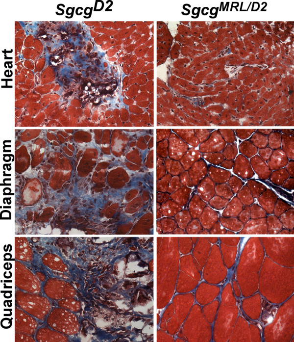
The MRL genome suppresses fibrosis in the heart and muscles of SgcgD2mice.Sgcg mice lack the dystrophin-associated protein, γ-sarcoglycan, and when in the DBA/2J (D2) background have a more severe phenotype with enhanced membrane leak and fibrosis [21]. A 50% contribution of the MRL genome, referred to as SgcgMRL/D2, suppressed fibrosis in both heart and skeletal muscle.
We quantified fibrosis by measuring HOP content as an indicator of collagen in SgcgD2 and SgcgMRL/D2 muscles. SgcgMRL/D2 mice have significantly reduced fibrosis compared to SgcgD2 mice (Figure (Figure2).2). We also evaluated fibrosis in aged (32 week) animals and found a persistent reduction in fibrosis in the SgcgMRL/D2 mice (Figure (Figure2,2, diagonal bars). The 129T2/SvEmsJ strain was previously shown to suppress the severity of muscle pathology in Sgcg null mice [33]. In comparison, the MRL background suppressed fibrosis more than the 129T2/SvEmsJ background (Figure (Figure3).3). The MRL background dramatically reduced fibrosis in the heart and abdominal muscles compared to 129T2/SvEmsJ. The MRL background also suppressed fibrosis more than the 129T2/SvEmsJ background in the diaphragm and quadriceps muscles, but to a lesser degree. This finding suggests that the phenotypically beneficial genetic modifiers in the MRL genome may be more potent than those in the 129T2/SvEmsJ genome.
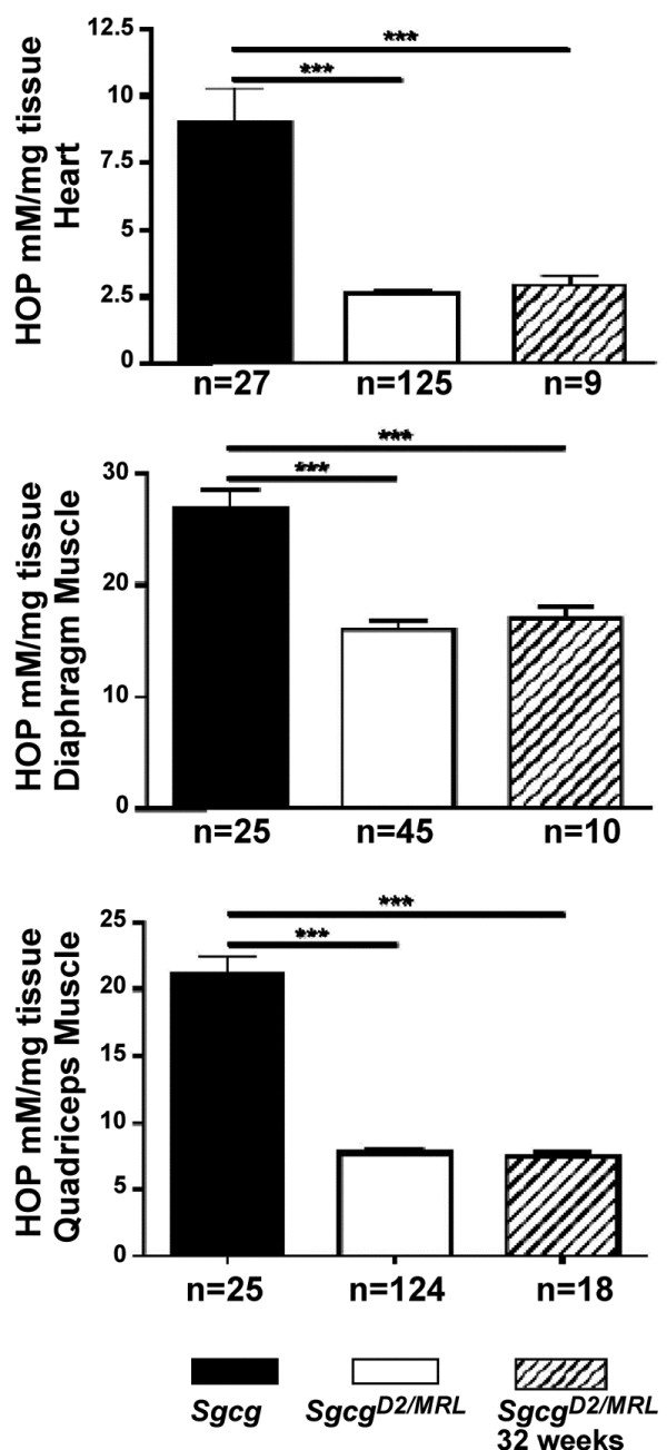
The MRL genome quantitatively reduces fibrosis in Sgcg mice. Hydroxyproline (HOP) is a measure of fibrosis and is represented on the y axis (mM/mg). In the heart, diaphragm and quadriceps muscles, fibrosis is significantly (p < 0.001) reduced in intercrossed SgcgMRL/D2 (D2/MRL) animals compared to Sgcg in the D2 (D2) background at 8 weeks. Fibrosis remains significantly (***p < 0.001) reduced in SgcgMRL/D2 animals at the 32-week time point.
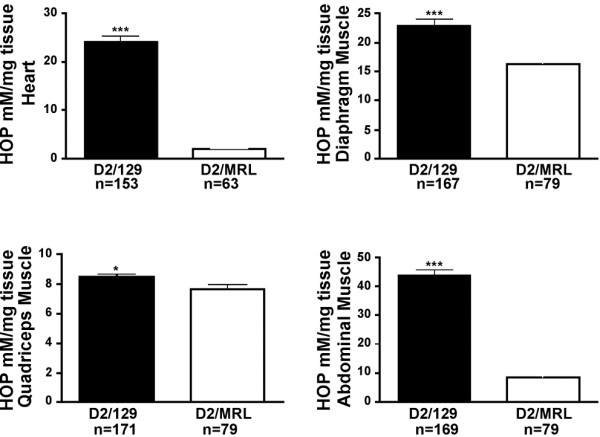
The reduction of fibrosis in intercrossed Sgcg animals is specific to the MRL genome. Hydroxyproline (HOP) is a measure of fibrosis and is represented on the y axis (mM/mg). Fibrosis is significantly reduced in the heart (***p < 0.001), diaphragm (p < 0.001), quadriceps (*p = 0.041) and abdominal muscles (p < 0.001) of Sgcg animals intercrossed in the D2/MRL background compared to those intercrossed in the D2/129 background.
The MRL background does not protect against membrane leak
Disruption of the membrane-associated dystrophin complex renders the sarcolemma unusually fragile, leading to abnormal membrane leakage that is visualized by uptake of the nonspecific vital tracer Evans blue dye [20]. Upon gross inspection, the muscles from the SgcgMRL/D2 mice displayed high levels of dye uptake, comparable to what was observed in the parental Sgcg mice (Additional file 1: Figure S1). On a microscopic level, dye-positive cells were readily detectable in SgcgMRL/D2 muscle and heart (Figure (Figure4).4). Evans blue dye levels were measured in multiple muscle groups and were not significantly different between SgcgMRL/D2 and SgcgD2 muscles for quadriceps, triceps, gastrocnemius/soleus, gluteus and abdominals muscle groups (Figure (Figure5).5). We also measured dye uptake in older animals at 32 weeks to assess disease progression. At 32 weeks, the skeletal muscles continued to show comparable levels of membrane leak as what was seen at 8 weeks (Figure (Figure5).5). The gluteus/hamstring group of muscles showed an increased of dye uptake compared to the 8-week animals. This increase was not seen for other muscle groups.
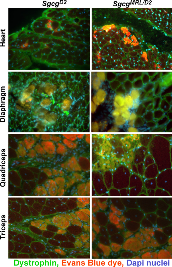
Membrane leak is not corrected by the presence of the MRL genome in Sgcg heart and muscle. Evans blue dye is found in cardiomyocytes and skeletal myofibers reflecting membrane leakiness. Dye uptake occurs in patchy pattern throughout the heart and muscle and is seen as opacified cells that fluoresce red. Nuclei are shown in blue and dystrophin in green.
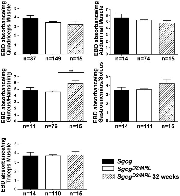
The MRL genome does not significantly reduce membrane damage in Sgcg muscle. Evans blue dye uptake is a measure of membrane damage and is represented on the y axis (absorbance/mg). In the quadriceps, triceps, abdominal and gastrocnemius/soleus muscles, membrane damage is not significantly different between SgcgD2 animals and intercrossed SgcgMRL/D2 animals at both the 8- and 32-week time points. In the gluteus/hamstring muscles, membrane damage is significantly (**p < 0.01) increased at the 32-week time point in intercrossed SgcgMRL/D2 animals compared to the 8-week time point.
We compared suppression of dye uptake by the MRL background to that induced by the 129T2/SvEmsJ background in Sgcg mice (Figure (Figure6).6). The quadriceps and abdominal muscles from Sgcg null mice with a contribution from the 129T2/SvEmsJ background showed increased membrane leak compared to Sgcg mice with an MRL contribution. However, the triceps muscle group from the MRL background showed increased dye uptake, and the gluteus/hamstring muscle group showed no significant difference. Thus, there was no consistent suppression of membrane leakiness by the MRL strain.
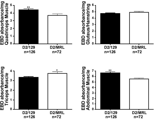
The MRL genome has a variable ability to reduce membrane leak in muscular dystrophy compared to the 129T2/SvEmsJ strain. Evans blue dye uptake was measured in multiple muscle groups represented on the y axis (absorbance/mg). Membrane damage is reduced in the quadriceps (**p = 0.0053) and abdominal (**p = 0.0012) muscles of intercrossed SgcgD2/MRL animals compared to intercrossed SgcgD2/129 mice. Membrane damage is increased in the triceps muscle (*p = 0.0298) of intercrossed SgcgMRL/D2 animals. There is no difference in membrane damage in the gluteus/hamstring muscle.
The MRL genome may promote skeletal muscle regeneration
The MRL background is thought to exert part of its effect by promoting regeneration [34]. We examined embryonic myosin heavy chain (eMHC) expression as a reflection of regeneration. SgcgMRL/D2 muscle shows patchy areas with a qualitative increase in eMHC-positive fibers compared to Sgcg (Figure (Figure7),7), and wild-type muscle showed no eMHC positive fibers (data not shown). In dystrophic skeletal muscle, ongoing regeneration is thought to offset degeneration. Consistent with this, eMHC-positive regions were also positive for Evans blue dye uptake, indicative of muscle damage. We also evaluated phosphorylated histone 3 (PH3) as a reflection of mitotic index. No clear differences for PH3 staining were seen between SgcgMRL/D2 and Sgcg muscle. Regenerating skeletal muscle, whether from trauma or muscular dystrophy, is identified by the presence of myofibers with centrally positioned nuclei. In muscular dystrophy, ongoing regeneration is also marked by increased fiber size variability. SgcgMRL/D2 mice have an increased number of central nuclei in quadriceps and diaphragm muscle compared to SgcgD2 mice (Figure (Figure7).7). These same muscles also showed increased fiber size variability when there is a contribution from the MRL background. These data are consistent with a model in which favorable matrix remodeling may support enhanced regeneration.
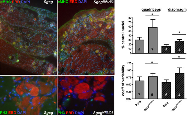
Regeneration may be enhanced in the MRL background. Embryonic myosin heavy chain (eMHC) staining was increased in SgcgMRL/D2 compared to Sgcg muscle, and eMHC fibers were seen in areas where dye uptake was seen. However, phosphorylated histone 3 (PH3), a marker of mitotic index, was not increased in SgcgMRL/D2 compared to Sgcg muscle. Skeletal muscle central nucleation and fiber size variability are enhanced by the MRL background. SgcgMRL/D2 muscle demonstrated an increase in centrally nucleated fibers compared to SgcgD2 mice. Fiber size variability was reflected in the coefficient of variability of fiber diameter measurements. The solid line indicates significance (p < 0.05). These data support that the MRL exerts its effect in skeletal muscle, at least in part, by promoting regeneration.
We also assessed apoptosis using a TUNEL assay and caspase 3 staining and again found no differences between SgcgMRL/D2 and Sgcg muscle, suggesting that gross differences in programmed cell death are unlikely to account for the improved healing of the MRL strain (Additional file 1: Figure S2). We also characterized whether T cell infiltration or macrophage infiltration differed qualitatively between SgcgMRL/D2 and Sgcg muscle, but we found no clear differences between SgcgMRL/D2 and Sgcg muscle, suggesting other features contribute to the MRL background’s effect on muscular dystrophy (Additional file 1: Figure S2).
Improved cardiac function from a 50% contribution of the MRL background
We performed 2D and M mode echocardiography on SgcgMRL/D2 and SgcgD2 mice at 12 weeks of age. Both fractional shortening and the left ventricular ejection fraction were significantly reduced in SgcgD2 mice, indicating that the increased fibrosis impairs cardiac function (Table (Table2).2). Fractional shortening and the left ventricular ejection fraction were similar between SgcgMRL/D2 and WTMRL/D2 mice, consistent with improved function mediated by 50% of the MRL genome. Wall thickness was also increased in Sgcg mice compared to the wild type of the same background. In contrast, wall thickness was not different between SgcgMRL/D2 and WTMRL/D2 mice.
Table 2
Cardiac function in Sgcg mice
| Fractional shortening | |
|---|---|
| SgcgD2(n = 4) | 33.87 ± 4.80a |
| WTD2 (n = 5) | 44.58 ± 7.65a |
| SgcgMRL/D2 (n = 5) | 46.00 ± 14.4 |
| WTMRL/D2 (n = 5) | 48.37 ± 11.21 |
ap = 0.046.
Chromosome 2 associates with reduced fibrosis Sgcg heart and diaphragm muscle
We conducted a genome-wide scan using markers that were informative in the two parental strains DBA/2J and MRL/MpJ (Figure (Figure8).8). QTLRel was used to identify regions of association; this analysis takes into account the relatedness of individual animals in the cohort [29,30]. A region on chromosome 2 was identified that associated with fibrosis in the heart. The 1.5-LOD drop interval of this region spans from 64.8008−73.1758 cM in mouse genome build 37.1. This same region was also associated with fibrosis in the diaphragm muscle and also for fibrosis in the abdominal muscles. It should be noted that the significance of these associations is suggestive (p < 0.63) when using the stringent QTLRel analysis. However, the overlapping intervals found in heart, diaphragm and abdominal muscles provide additional support that this interval modifies fibrosis.
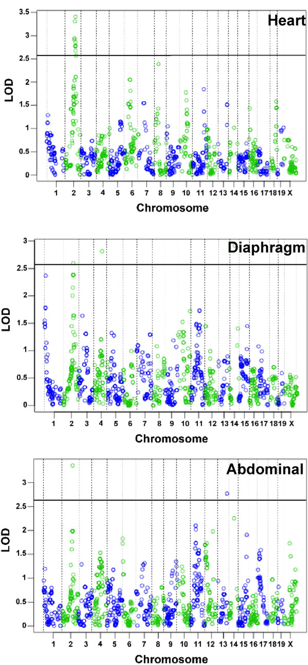
Fibrosis in the heart, diaphragm and abdominal muscles is modified by a locus on chromosome 2. Genome-wide association was examined for fibrosis in SgcgMRL/D2 progeny in the (A) cardiac muscle (n = 65), (B) diaphragm (n = 78) and (C) abdominal muscle (n = 78). Chromosomes are plotted on the x axis, and the informative SNPs tested (n = 1,707) are displayed as open circles that alternate color by chromosome; LOD scores are represented on the y axis. Overlapping regions on chromosome 2 showed suggestive (p < 0.63) association with fibrosis in heart, diaphragm and abdominal muscle.
Ltbp4 polymorphism does not account for the MRL healing properties
The DBA/2J strain contains an insertion/deletion polymorphism within the Ltbp4 gene [21] that modifies both membrane permeability and fibrosis, as two independent traits, in Sgcg muscular dystrophy. Ltbp4 encodes latent TGFβ-binding protein 4, and TGFβ proteins have been extensively linked to fibrosis in many disease states including muscular dystrophy [35,36]. Most murine strains, including the MRL/MpJ strain used here, contain the protective Ltbp4 allele with an additional 12 amino acids inserted in exon 12. Because the SgcgMRL/D2 cohort used here contained both the protective (LtbpI allele) and the disease-enhancing allele (Ltbp4D), we tested whether Ltbp4 genotype correlated with fibrosis in the SgcgMRL/D2 cohort. Table Table33 shows that neither membrane permeability nor fibrosis is correlated with the Ltbp4 genotype in the SgcgMRL/DBA2J cohort. Therefore, Ltbp4 does not account for the suppressive effect of the MRL background, and other genetic modifiers account for this difference.
Table 3
Ltbp4 genotype in SgcgMRL/D2mice
| Dye uptake | Fibrosis | |
|---|---|---|
| Ltbp4D/D (n = 14) | 3.90 ± 0.83 | 6.00 ± 1.81 |
| Ltbp4D/I (n = 34) | 4.18 ± 0.84 | 8.05 ± 3.02 |
| LtbpI/I (n = 47) | 4.00 ± 0.85 | 6.64 ± 2.15 |
Discussion
The MRL genome protects against fibrosis but not membrane leak
Mutations in the dystrophin or sarcoglycan genes share a common pathological mechanism characterized by disruption of the plasma membrane of cardiomyocytes and skeletal myofibers. Membrane leak is an early step in the pathological process while fibrosis is thought to be a secondary response. Our study suggests that there are genetic pathways that target fibrosis without altering the sarcolemmal leak properties. In the heart, reduced fibrosis was correlated with functional benefit, although cardiac function may be improved from both cardiac intrinsic as well as cardiac extrinsic features. For example, improved skeletal muscle function may contribute to improved cardiac function, particularly when considering the contribution of the respiratory musculature.
The MRL strain typically shows the improved healing capability in its first 2–6 months of life. After 6 months of age, wild-type MRL mice develop autoimmune disorders; it is for this reason that we conducted studies using a 50% contribution of the MRL strain. Quantitative trait mapping has been used to define many different genetic regions associated with the healing properties [14]. The autoimmune properties have been genetically separated from the healing properties since the four to six different “autoimmune” genetic loci do not overlap with those associated with improved healing. In our studies, only 50% of the MRL genome was capable of suppressing fibrosis and improving function. In the course of these studies, we never identified a single SgcgMRL/D2 animal that had markedly elevated fibrosis. This stands in contrast to what is normally observed in the Sgcg animals where outliers with markedly elevated fibrosis are often seen. The maximum values for fibrosis in SgcgMRL/D2 mice were all well below what was measured in SgcgD2 mice. This is consistent with multiple, dominant genetic loci imparting the improved healing capabilities, similar to what has been observed for the ear hole repair properties in the MRL strain [37]. The finding that improvement persists in the SgcgMRL/D2 mice, with almost complete suppression of fibrosis at 32 weeks, suggests that a persistent healing effect can arise from the MRL strain.
Chromosome 2 candidate genes
When considering the overlapping region on chromosome 2 from the heart, diaphragm and abdominal fibrosis data, the interval contains 49 known genes. There are additional predicted and unnamed genes and genes encoding olfactory receptors that are not considered likely candidates. Within the interval is Jag1, encoding jagged the ligand for the Notch receptor. Loss of function of Notch 3 leads to muscle hyperplasia, especially when subjected to repetitive injury [38]. Ex vivo activation of Notch signaling helps maintain donor cell engraftment during myoblast transfer [39]. Another gene linked to growth and healing in the interval is Bmp2-encoding bone morphogenetic protein 2. The BMPs mediate musculoskeletal regeneration [40], and given the MRL background effect on multiple cells and tissues, BMP2 is well positioned to contribute to the MRL superhealing response. That said, this chromosome 2 interval has not previously been linked to other MRL healing properties. The strict criteria imposed by the QTLRel algorithm makes it unlikely that these results derive from relatedness of animals within the cohort.
Cardiac injury and repair in the MRL strain
The ability of the heart to recover after injury has been studied in the MRL strain using cryoinjury, left anterior descending ligation and ischemia reperfusion methods [5,6,8-10,14,15]. However, scar reduction has been noted after some forms of injury while not after others. In common to all these studies is an acute injury model where a normal heart was substantially damaged in a single setting. The MRL’s ability to heal the heart may be limited such that larger amounts of injury may be insurmountable, as proposed by Naseem [6]. Our data support that lower, although persistent, levels of injury can be managed by the MRL strain where there is significant reduction in fibrosis and a corresponding functional improvement in cardiac function.
Mechanisms for MRL healing
A number of mechanisms likely act in concert to achieve suppression of fibrosis and improvement of function. In skeletal muscle, where muscle stem cells robustly regenerate muscle after injury, there is indirect evidence of enhanced regeneration. Specifically, there is an increase in centrally nucleated myofibers, which is thought to reflect enhanced myoblast fusion. However, our data do not distinguish whether the MRL background exerts its effect on muscle regenerative cells, or by creating a more supportive extracellular matrix or both. MRL animals heal with embryonic characteristics with enhanced blastema formation and metabolic and gene expression features consistent with an earlier developmental state [41]. Altered protease expression has also been noted in the MRL mouse in response to damage [5]. Recent work characterizing the immune infiltrate in muscular dystrophy found that reducing osteopontin was effective at suppressing fibrosis [17]. Together these data may favor matrix-associated modifications that can modulate muscle fibrosis and function.
Conclusions
Herein the MRL background was shown to reduce fibrosis in a chronic model of muscular dystrophy and cardiomyopathy, the Sgcg mouse. Although membrane leak was still evident in Sgcg mice sharing a portion of the MRL genome, the reduction in fibrosis was associated with improved cardiac function. The identification of gene(s) from the MRL genome will help identify pathways important for chronic repair of myopathic processes.
Abbreviations
BMP: Bone morphogenetic protein; D2: (DBA/2J); DMD: Duchenne muscular dystrophy; HOP: Hydroxyproline; LTBP4: Latent TGFβ-binding protein; MRL: Murphy Roth Large; QTL: Quantitative trait loci; SNP: Single nucleotide polymorphism; TGFβ: Transforming growth factor β.
Competing interest
The authors have no competing interests related to this work.
Authors’ contributions
AH conducted the phenotypic analysis of muscle. KAS conducted the genome-wide SNP analysis and phenotype analysis. GK performed the echocardiographic analysis. MH oversaw the breeding. JHC assisted with phenotypic analysis. EMM conceived the experiments, analyzed the data and wrote the manuscript. All authors read and approved the final manuscript.
Supplementary Material
Figure S1. Shown are gross images from Sgcg vs. SgcgMRL/D2 mice. Evans blue dye uptake could be readily seen in the quadriceps and diaphragm muscles and did not appear grossly altered by the presence of the MRL background. In contrast, fibrosis was visually reduced in the quadriceps and heart of SgcgMRL/D2 compared to Sgcg mice. The diaphragm muscle retained evidence of fibrosis in SgcgMRL/D2, but the white stripes of fibrosis were smaller, and intact diaphragm muscle was still evident compared to the near total replacement of diaphragm muscle in Sgcg mice. Figure S2. Shown is staining for apoptosis with TUNEL and caspase indicating no gross differences between Sgcg and SgcgMRL/D2 muscle. CD3 and MAC1 staining to examine T cell and macrophage infiltrate also did not appear grossly altered by the presence of the MRL background.
Acknowledgements
Supported by NIH R01HL61322 (EMM), R01HL102322 (AH) and NIH K08HL098565 (GK).
References
- Clark LD, Clark RK, Heber-Katz E. A new murine model for mammalian wound repair and regeneration. Clin Immunol Immunopathol. 1998;88:35–45. 10.1006/clin.1998.4519. [Abstract] [CrossRef] [Google Scholar]
- McBrearty BA, Clark LD, Zhang XM, Blankenhorn EP, Heber-Katz E. Genetic analysis of a mammalian wound-healing trait. Proc Natl Acad Sci U S A. 1998;95:11792–11797. 10.1073/pnas.95.20.11792. [Europe PMC free article] [Abstract] [CrossRef] [Google Scholar]
- Gourevitch DL, Clark L, Bedelbaeva K, Leferovich J, Heber-Katz E. Dynamic changes after murine digit amputation: the MRL mouse digit shows waves of tissue remodeling, growth, and apoptosis. Wound Repair Regen. 2009;17:447–455. 10.1111/j.1524-475X.2009.00492.x. [Europe PMC free article] [Abstract] [CrossRef] [Google Scholar]
- Ueno M, Lyons BL, Burzenski LM, Gott B, Shaffer DJ, Roopenian DC, Shultz LD. Accelerated wound healing of alkali-burned corneas in MRL mice is associated with a reduced inflammatory signature. Invest Ophthalmol Vis Sci. 2005;46:4097–4106. 10.1167/iovs.05-0548. [Abstract] [CrossRef] [Google Scholar]
- Leferovich JM, Bedelbaeva K, Samulewicz S, Zhang XM, Zwas D, Lankford EB, Heber-Katz E. Heart regeneration in adult MRL mice. Proc Natl Acad Sci U S A. 2001;98:9830–9835. 10.1073/pnas.181329398. [Europe PMC free article] [Abstract] [CrossRef] [Google Scholar]
- Naseem RH, Meeson AP, Michael Dimaio J, White MD, Kallhoff J, Humphries C, Goetsch SC, De Windt LJ, Williams MA, Garry MG, Garry DJ. Reparative myocardial mechanisms in adult C57BL/6 and MRL mice following injury. Physiol Genomics. 2007;30:44–52. 10.1152/physiolgenomics.00070.2006. [Abstract] [CrossRef] [Google Scholar]
- Abdullah I, Lepore JJ, Epstein JA, Parmacek MS, Gruber PJ. MRL mice fail to heal the heart in response to ischemia-reperfusion injury. Wound Repair Regen. 2005;13:205–208. 10.1111/j.1067-1927.2005.130212.x. [Abstract] [CrossRef] [Google Scholar]
- Cimini M, Fazel S, Fujii H, Zhou S, Tang G, Weisel RD, Li RK. The MRL mouse heart does not recover ventricular function after a myocardial infarction. Cardiovasc Pathol. 2008;17:32–39. 10.1016/j.carpath.2007.06.006. [Abstract] [CrossRef] [Google Scholar]
- Grisel P, Meinhardt A, Lehr HA, Kappenberger L, Barrandon Y, Vassalli G. The MRL mouse repairs both cryogenic and ischemic myocardial infarcts with scar. Cardiovasc Pathol. 2008;17:14–22. 10.1016/j.carpath.2007.01.007. [Abstract] [CrossRef] [Google Scholar]
- Robey TE, Murry CE. Absence of regeneration in the MRL/MpJ mouse heart following infarction or cryoinjury. Cardiovasc Pathol. 2008;17:6–13. 10.1016/j.carpath.2007.01.005. [Europe PMC free article] [Abstract] [CrossRef] [Google Scholar]
- Oh YS, Thomson LE, Fishbein MC, Berman DS, Sharifi B, Chen PS. Scar formation after ischemic myocardial injury in MRL mice. Cardiovasc Pathol. 2004;13:203–206. 10.1016/j.carpath.2004.03.610. [Abstract] [CrossRef] [Google Scholar]
- Harty M, Neff AW, King MW, Mescher AL. Regeneration or scarring: an immunologic perspective. Dev Dyn. 2003;226:268–279. 10.1002/dvdy.10239. [Abstract] [CrossRef] [Google Scholar]
- Gourevitch D, Clark L, Chen P, Seitz A, Samulewicz SJ, Heber-Katz E. Matrix metalloproteinase activity correlates with blastema formation in the regenerating MRL mouse ear hole model. Dev Dyn. 2003;226:377–387. 10.1002/dvdy.10243. [Abstract] [CrossRef] [Google Scholar]
- Heber-Katz E, Leferovich J, Bedelbaeva K, Gourevitch D, Clark L. The scarless heart and the MRL mouse. Philos Trans R Soc Lond B Biol Sci. 2004;359:785–793. 10.1098/rstb.2004.1468. [Europe PMC free article] [Abstract] [CrossRef] [Google Scholar]
- Bedelbaeva K, Gourevitch D, Clark L, Chen P, Leferovich JM, Heber-Katz E. The MRL mouse heart healing response shows donor dominance in allogeneic fetal liver chimeric mice. Cloning Stem Cells. 2004;6:352–363. 10.1089/clo.2004.6.352. [Abstract] [CrossRef] [Google Scholar]
- Heydemann A. The super super-healing MRL mouse strain. Front Biol. 2012. in press. [Europe PMC free article] [Abstract]
- Spencer MJ, Montecino-Rodriguez E, Dorshkind K, Tidball JG. Helper (CD4(+)) and cytotoxic (CD8(+)) T cells promote the pathology of dystrophin-deficient muscle. Clin Immunol. 2001;98:235–243. 10.1006/clim.2000.4966. [Abstract] [CrossRef] [Google Scholar]
- Hack AA, Ly CT, Jiang F, Clendenin CJ, Sigrist KS, Wollmann RL, McNally EM. Gamma-sarcoglycan deficiency leads to muscle membrane defects and apoptosis independent of dystrophin. J Cell Biol. 1998;142:1279–1287. 10.1083/jcb.142.5.1279. [Europe PMC free article] [Abstract] [CrossRef] [Google Scholar]
- Heydemann A, Huber JM, Demonbreun A, Hadhazy M, McNally EM. Genetic background influences muscular dystrophy. Neuromuscul Disord. 2005;15:601–609. 10.1016/j.nmd.2005.05.004. [Abstract] [CrossRef] [Google Scholar]
- Straub V, Rafael JA, Chamberlain JS, Campbell KP. Animal models for muscular dystrophy show different patterns of sarcolemmal disruption. J Cell Biol. 1997;139:375–385. 10.1083/jcb.139.2.375. [Europe PMC free article] [Abstract] [CrossRef] [Google Scholar]
- Heydemann A, Ceco E, Lim JE, Hadhazy M, Ryder P, Moran JL, Beier DR, Palmer AA, McNally EM. Latent TGF-beta-binding protein 4 modifies muscular dystrophy in mice. J Clin Invest. 2009;119:3703–3712. 10.1172/JCI39845. [Europe PMC free article] [Abstract] [CrossRef] [Google Scholar]
- Matsuda R, Nishikawa A, Tanaka H. Visualization of dystrophic muscle fibers in mdx mouse by vital staining with Evans blue: evidence of apoptosis in dystrophin-deficient muscle. J Biochem (Tokyo) 1995;118:959–964. 10.1093/jb/118.5.959. [Abstract] [CrossRef] [Google Scholar]
- Thurston G, Suri C, Smith K, McClain J, Sato TN, Yancopoulos GD, McDonald DM. Leakage-resistant blood vessels in mice transgenically overexpressing angiopoietin-1. Science. 1999;286:2511–2514. 10.1126/science.286.5449.2511. [Abstract] [CrossRef] [Google Scholar]
- Flesch M, Schiffer F, Zolk O, Pinto Y, Rosenkranz S, Hirth-Dietrich C, Arnold G, Paul M, Bohm M. Contractile systolic and diastolic dysfunction in renin-induced hypertensive cardiomyopathy. Hypertension. 1997;30:383–391. 10.1161/01.HYP.30.3.383. [Abstract] [CrossRef] [Google Scholar]
- Collins KA, Korcarz CE, Shroff SG, Bednarz JE, Fentzke RC, Lin H, Leiden JM, Lang RM. Accuracy of echocardiographic estimates of left ventricular mass in mice. Am J Physiol Heart Circ Physiol. 2001;280:H1954–H1962. [Abstract] [Google Scholar]
- Wheeler MT, Korcarz CE, Collins KA, Lapidos KA, Hack AA, Lyons MR, Zarnegar S, Earley JU, Lang RM, McNally EM. Secondary coronary artery vasospasm promotes cardiomyopathy progression. Am J Pathol. 2004;164:1063–1071. 10.1016/S0002-9440(10)63193-8. [Europe PMC free article] [Abstract] [CrossRef] [Google Scholar]
- Moran JL, Bolton AD, Tran PV, Brown A, Dwyer ND, Manning DK, Bjork BC, Li C, Montgomery K, Siepka SM, Vitaterna MH, Takahashi JS, Wiltshire T, Kwiatkowski DJ, Kucherlapati R, Beier DR. Utilization of a whole genome SNP panel for efficient genetic mapping in the mouse. Genome Res. 2006;16:436–440. 10.1101/gr.4563306. [Europe PMC free article] [Abstract] [CrossRef] [Google Scholar]
- Collaborative Cross Consortium. The genome architecture of the Collaborative Cross mouse genetic reference population. Genetics. 2012;190:389–401. [Europe PMC free article] [Abstract] [Google Scholar]
- Cheng R, Abney M, Palmer AA, Skol AD. QTLRel: an R package for genome-wide association studies in which relatedness is a concern. BMC Genet. 2011;12:66. [Europe PMC free article] [Abstract] [Google Scholar]
- Cheng R, Lim JE, Samocha KE, Sokoloff G, Abney M, Skol AD, Palmer AA. Genome-wide association studies and the problem of relatedness among advanced intercross lines and other highly recombinant populations. Genetics. 2010;185:1033–1044. 10.1534/genetics.110.116863. [Europe PMC free article] [Abstract] [CrossRef] [Google Scholar]
- McNally EM, Passos-Bueno MR, Bonnemann CG, Vainzof M, de Sa Moreira E, Lidov HG, Othmane KB, Denton PH, Vance JM, Zatz M, Kunkel LM. Mild and severe muscular dystrophy caused by a single gamma-sarcoglycan mutation. Am J Hum Genet. 1996;59:1040–1047. [Europe PMC free article] [Abstract] [Google Scholar]
- Stedman HH, Sweeney HL, Shrager JB, Maguire HC, Panettieri RA, Petrof B, Narusawa M, Leferovich JM, Sladky JT, Kelly AM. The mdx mouse diaphragm reproduces the degenerative changes of Duchenne muscular dystrophy. Nature. 1991;352:536–539. 10.1038/352536a0. [Abstract] [CrossRef] [Google Scholar]
- Swaggart KA, Heydemann A, Palmer AA, McNally EM. Distinct genetic regions modify specific muscle groups in muscular dystrophy. Physiol Genomics. 2011;43:24–31. 10.1152/physiolgenomics.00172.2010. [Europe PMC free article] [Abstract] [CrossRef] [Google Scholar]
- Bedelbaeva K, Snyder A, Gourevitch D, Clark L, Zhang XM, Leferovich J, Cheverud JM, Lieberman P, Heber-Katz E. Lack of p21 expression links cell cycle control and appendage regeneration in mice. Proc Natl Acad Sci U S A. 2010;107:5845–5850. 10.1073/pnas.1000830107. [Europe PMC free article] [Abstract] [CrossRef] [Google Scholar]
- Bernasconi P, Torchiana E, Confalonieri P, Brugnoni R, Barresi R, Mora M, Cornelio F, Morandi L, Mantegazza R. Expression of transforming growth factor-beta 1 in dystrophic patient muscles correlates with fibrosis. Pathogenetic role of a fibrogenic cytokine. J Clin Invest. 1995;96:1137–1144. 10.1172/JCI118101. [Europe PMC free article] [Abstract] [CrossRef] [Google Scholar]
- Cohn RD, Van Erp C, Habashi JP, Soleimani AA, Klein EC, Lisi MT, Gamradt M, ap Rhys CM, Holm TM, Loeys BL, Ramirez F, Judge DP, Ward CW, Dietz HC. Angiotensin II type 1 receptor blockade attenuates TGF-beta-induced failure of muscle regeneration in multiple myopathic states. Nat Med. 2007;13:204–210. 10.1038/nm1536. [Europe PMC free article] [Abstract] [CrossRef] [Google Scholar]
- Kench JA, Russell DM, Fadok VA, Young SK, Worthen GS, Jones-Carson J, Henson JE, Henson PM, Nemazee D. Aberrant wound healing and TGF-beta production in the autoimmune-prone MRL/+ mouse. Clin Immunol. 1999;92:300–310. 10.1006/clim.1999.4754. [Abstract] [CrossRef] [Google Scholar]
- Kitamoto T, Hanaoka K. Notch3 null mutation in mice causes muscle hyperplasia by repetitive muscle regeneration. Stem Cells. 2010;28:2205–2216. 10.1002/stem.547. [Abstract] [CrossRef] [Google Scholar]
- Parker MH, Loretz C, Tyler AE, Duddy WJ, Hall JK, Olwin BB, Bernstein ID, Storb R, Tapscott SJ. Activation of notch signaling during ex vivo expansion maintains donor muscle cell engraftment. Stem Cells. 2012;30:2212–20. 10.1002/stem.1181. [Europe PMC free article] [Abstract] [CrossRef] [Google Scholar]
- Ruschke K, Hiepen C, Becker J, Knaus P. BMPs are mediators in tissue crosstalk of the regenerating musculoskeletal system. Cell Tissue Res. 2012;347:521–544. 10.1007/s00441-011-1283-6. [Abstract] [CrossRef] [Google Scholar]
- Naviaux RK, Le TP, Bedelbaeva K, Leferovich J, Gourevitch D, Sachadyn P, Zhang XM, Clark L, Heber-Katz E. Retained features of embryonic metabolism in the adult MRL mouse. Mol Genet Metab. 2009;96:133–144. 10.1016/j.ymgme.2008.11.164. [Europe PMC free article] [Abstract] [CrossRef] [Google Scholar]
Articles from Skeletal Muscle are provided here courtesy of BMC
Full text links
Read article at publisher's site: https://doi.org/10.1186/2044-5040-2-26
Read article for free, from open access legal sources, via Unpaywall:
https://skeletalmusclejournal.biomedcentral.com/track/pdf/10.1186/2044-5040-2-26
Citations & impact
Impact metrics
Article citations
The super-healing MRL strain promotes muscle growth in muscular dystrophy through a regenerative extracellular matrix.
JCI Insight, 9(3):e173246, 04 Jan 2024
Cited by: 2 articles | PMID: 38175727 | PMCID: PMC11143963
Altered TGFB1 regulated pathways promote accelerated tendon healing in the superhealer MRL/MpJ mouse.
Sci Rep, 12(1):3026, 22 Feb 2022
Cited by: 9 articles | PMID: 35194136 | PMCID: PMC8863792
The MRL/MpJ Mouse Strain Is Not Protected From Muscle Atrophy and Weakness After Rotator Cuff Tear.
J Orthop Res, 38(4):811-822, 17 Nov 2019
Cited by: 2 articles | PMID: 31696955 | PMCID: PMC7071998
Laminin-111 protein therapy enhances muscle regeneration and repair in the GRMD dog model of Duchenne muscular dystrophy.
Hum Mol Genet, 28(16):2686-2695, 01 Aug 2019
Cited by: 15 articles | PMID: 31179490 | PMCID: PMC6687953
Sunitinib promotes myogenic regeneration and mitigates disease progression in the mdx mouse model of Duchenne muscular dystrophy.
Hum Mol Genet, 28(13):2120-2132, 01 Jul 2019
Cited by: 7 articles | PMID: 30806670 | PMCID: PMC6586148
Go to all (25) article citations
Other citations
Data
Data behind the article
This data has been text mined from the article, or deposited into data resources.
BioStudies: supplemental material and supporting data
Similar Articles
To arrive at the top five similar articles we use a word-weighted algorithm to compare words from the Title and Abstract of each citation.
Cardiac function in muscular dystrophy associates with abdominal muscle pathology.
J Neuromuscul Dis, 2(1):39-49, 01 Jan 2015
Cited by: 3 articles | PMID: 26029630 | PMCID: PMC4447317
Distinct genetic regions modify specific muscle groups in muscular dystrophy.
Physiol Genomics, 43(1):24-31, 19 Oct 2010
Cited by: 23 articles | PMID: 20959497 | PMCID: PMC3026347
The super-healing MRL strain promotes muscle growth in muscular dystrophy through a regenerative extracellular matrix.
JCI Insight, 9(3):e173246, 04 Jan 2024
Cited by: 2 articles | PMID: 38175727 | PMCID: PMC11143963
Modifiers of heart and muscle function: where genetics meets physiology.
Exp Physiol, 99(4):621-626, 08 Nov 2013
Cited by: 15 articles | PMID: 24213858 | PMCID: PMC3975685
Review Free full text in Europe PMC
Funding
Funders who supported this work.
NHLBI NIH HHS (1)
Grant ID: R01 HL102322
NIAMS NIH HHS (1)
Grant ID: U54 AR052646

 1,2
1,2


