| PMC full text: | Published online 2016 Mar 15. doi: 10.1371/journal.pone.0151589
|
Fig 5

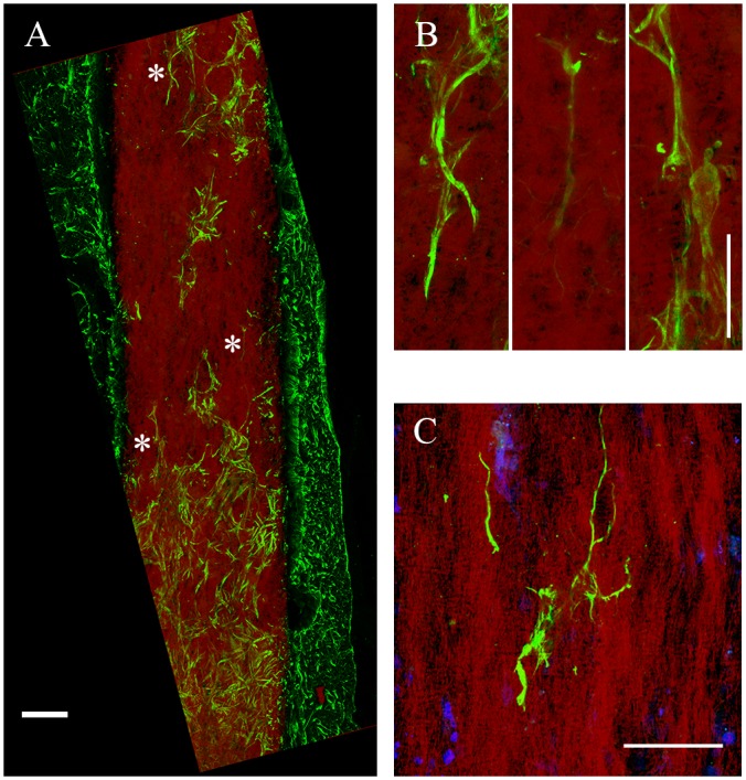
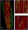
Astrocyte directional growth with a gP6 scaffold following process infiltration.
(A) astrocytes and processes change their cell/process alignment to the microfiber direction to follow fiber alignment after infiltration into gP6 implant. (B) Enlarged areas in (A, marked by *) indicating astrocytic processes alignment along the vertical axis. (C) Multiple process alignment of a single astrocyte within the scaffold. Brain tissue sections were collected in the sagittal plane. Scale bars in (A) and (B, C) represent 100 and 20 μm respectively.


