Logo of phersnaLink to Publisher's site
PMC full text:

Figure 5.

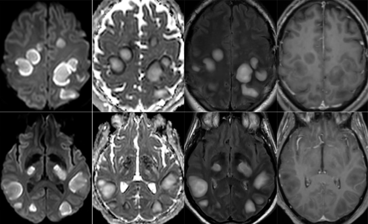
54-year old man with pathological wakefulness after sedation. Non-confluent multifocal white matter hyperintense lesions on FLAIR and diffusion, with variable enhancement. Axial Diffusion (A, B), Apparent Diffusion Coefficient (ADC) map (C, D), axial postcontrast FLAIR (E, F), and postcontrast T1 weighted MR images (G, H). Multiple nodular hyperintense Diffusion and FLAIR subcortical and corticospinal tracts lesions, with very mild mass effect on adjacent structures. The lesions present a center with an elevation of ADC corresponding to vasogenic edema and a peripheral ring of reduced ADC corresponding to cytotoxic edema (C, D). After contrast administration, small areas of very mild enhancement are detected (G, H).
54-year old man with pathological wakefulness after sedation. Non-confluent multifocal white matter hyperintense lesions on FLAIR and diffusion, with variable enhancement. Axial Diffusion (A, B), Apparent Diffusion Coefficient (ADC) map (C, D), axial postcontrast FLAIR (E, F), and postcontrast T1 weighted MR images (G, H). Multiple nodular hyperintense Diffusion and FLAIR subcortical and corticospinal tracts lesions, with very mild mass effect on adjacent structures. The lesions present a center with an elevation of ADC corresponding to vasogenic edema and a peripheral ring of reduced ADC corresponding to cytotoxic edema (C, D). After contrast administration, small areas of very mild enhancement are detected (G, H).

Images in this article

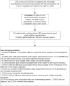
  • Flowchart of patient inclusion and exclusion
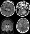
  • Axial FLAIR in four different COVID-19 patients. A) 58-year old man with impaired consciousness: FLAIR hyperintensities located in the left medial temporal lobe. B) 66-year old man with impaired consciousness: FLAIR ovoid hyperintense lesion located in the central part of the splenium of the corpus callosum. C) 71-year old woman with pathological wakefulness after sedation: extensive and confluent supratentorial white matter FLAIR hyperintensities (arrows). Association with leptomeningeal enhancement (stars) D) 61-year old man with confusion: hyperintense lesions involving both middle cerebellar peduncles.
  • 65-year old man with pathological wakefulness after sedation. Non-confluent multifocal white matter hyperintense lesions on FLAIR and diffusion, with variable enhancement, and hemorrhagic lesions. Axial Diffusion (A, B), Apparent Diffusion Coefficient (ADC) map (C), axial FLAIR (D, E), sagittal FLAIR (F), axial Susceptibility weighted imaging (SWI) (G), and postcontrast T1 weighted MR images (H). Multiple nodular hyperintense Diffusion and FLAIR lesions localized in the white matter including the corpus callosum (F). Some of them (white arrow) are associated with reduced ADC corresponding to cytotoxic edema (C). Other lesions are located next to the lenticular nucleus (cross) (E, G, H), with hemorrhagic changes (G), and enhancement after contrast administration.
  • 57-year old man with pathological wakefulness after sedation. Extensive and isolated white matter microhemorrhages. Axial Susceptibility weighted imaging (SWI) (A, B, C, D): multiple microhemorrhages mainly affecting the subcortical white matter, corpus callosum, internal capsule, and cerebellar peduncles.
  • 54-year old man with pathological wakefulness after sedation. Non-confluent multifocal white matter hyperintense lesions on FLAIR and diffusion, with variable enhancement. Axial Diffusion (A, B), Apparent Diffusion Coefficient (ADC) map (C, D), axial postcontrast FLAIR (E, F), and postcontrast T1 weighted MR images (G, H). Multiple nodular hyperintense Diffusion and FLAIR subcortical and corticospinal tracts lesions, with very mild mass effect on adjacent structures. The lesions present a center with an elevation of ADC corresponding to vasogenic edema and a peripheral ring of reduced ADC corresponding to cytotoxic edema (C, D). After contrast administration, small areas of very mild enhancement are detected (G, H).
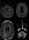
  • 51-year old man with impaired consciousness. Acute necrotizing encephalopathy. Axial FLAIR (A, C, D), and coronal FLAIR (B): bilateral FLAIR hyperintensity (cross) in both thalami (A, B), associated with involvement of the cerebellar (C), and cerebral (D) white matter (arrows).
Click on the image to see a larger version.