Abstract
Free full text

Analysis of SARS-CoV-2 RNA Persistence across Indoor Surface Materials Reveals Best Practices for Environmental Monitoring Programs
ABSTRACT
Environmental monitoring in public spaces can be used to identify surfaces contaminated by persons with coronavirus disease 2019 (COVID-19) and inform appropriate infection mitigation responses. Research groups have reported detection of severe acute respiratory syndrome coronavirus 2 (SARS-CoV-2) on surfaces days or weeks after the virus has been deposited, making it difficult to estimate when an infected individual may have shed virus onto a SARS-CoV-2-positive surface, which in turn complicates the process of establishing effective quarantine measures. In this study, we determined that reverse transcription-quantitative PCR (RT-qPCR) detection of viral RNA from heat-inactivated particles experiences minimal decay over 7 days of monitoring on eight out of nine surfaces tested. The properties of the studied surfaces result in RT-qPCR signatures that can be segregated into two material categories, rough and smooth, where smooth surfaces have a lower limit of detection. RT-qPCR signal intensity (average quantification cycle [Cq]) can be correlated with surface viral load using only one linear regression model per material category. The same experiment was performed with untreated viral particles on one surface from each category, with essentially identical results. The stability of RT-qPCR viral signal demonstrates the need to clean monitored surfaces after sampling to establish temporal resolution. Additionally, these findings can be used to minimize the number of materials and time points tested and allow for the use of heat-inactivated viral particles when optimizing environmental monitoring methods.
IMPORTANCE Environmental monitoring is an important tool for public health surveillance, particularly in settings with low rates of diagnostic testing. Time between sampling public environments, such as hospitals or schools, and notifying stakeholders of the results should be minimal, allowing decisions to be made toward containing outbreaks of coronavirus disease 2019 (COVID-19). The Safer At School Early Alert program (SASEA) (https://saseasystem.org/), a large-scale environmental monitoring effort in elementary school and child care settings, has processed >13,000 surface samples for SARS-CoV-2, detecting viral signals from 574 samples. However, consecutive detection events necessitated the present study to establish appropriate response practices around persistent viral signals on classroom surfaces. Other research groups and clinical labs developing environmental monitoring methods may need to establish their own correlation between RT-qPCR results and viral load, but this work provides evidence justifying simplified experimental designs, like reduced testing materials and the use of heat-inactivated viral particles.
OBSERVATION
Development and characterization of methods for environmental monitoring of severe acute respiratory syndrome coronavirus 2 (SARS-CoV-2) remain important areas of research for identifying and mitigating potential outbreaks as the global pandemic continues. Environmental monitoring offers indirect detection of possibly infectious individuals through noninvasive sampling. In spaces with relatively consistent occupants, detection of SARS-CoV-2 from environmental samples can help identify coronavirus disease 2019 (COVID-19)-infected individuals, ideally before further transmission. Environmental monitoring can also alert public health leadership to the potential presence of an infection even in settings with low diagnostic testing uptake, allowing for the implementation of enhanced nonpharmaceutical interventions (i.e., double masking, increased hand hygiene, improved ventilation efforts) even in the absence of positive diagnostic tests.
SARS-CoV-2 particles are shed by symptomatic and asymptomatic carriers (1) and have been detected on various surfaces (2,–5). Viral signatures have been demonstrated to persist up to 4 weeks in bulk floor dust collected from a room with a quarantined individual (6). Previous environmental monitoring studies have detected SARS-CoV-2 on surfaces contaminated by infected individuals in hospitals and congregate care facilities (6,–10). Thus, indoor surface sampling can be valuable for detection of infected persons indoors, where transmission risk is highest (11). The Safer At School Early Alert program (SASEA) (https://saseasystem.org/) uses environmental monitoring and collected over 13,000 surface swabs, but we need more information to clarify what these data are telling us over time.
We sought to characterize temporal dynamics underlying detection of SARS-CoV-2 signals from surface swabs from a variety of common indoor surface types using reverse transcription-quantitative PCR (RT-qPCR). The Centers for Disease Control and Prevention (CDC) maintains that the risk of fomite transmission of SARS-CoV-2 is low (12). This study makes no claims of attempting to understand the possibility of or mechanisms behind infection of virus transmitted by fomites but rather on whether and how negative and positive RT-qPCR detection from surface swabs can enable decision-making in outbreak mitigation, focused clinical testing of individuals, and safe reopening of high-traffic, public spaces.
We used RT-qPCR to detect heat-inactivated viral particles on nine surface materials and monitored the persistence of the heat-inactivated virus for 7 days. Each material—acrylic, steel, glass, ceramic tile, melamine-finished particleboard (MFP), painted drywall, vinyl flooring, and two different carpets (olefin and polyester)—was divided into 5-cm by 5-cm grids, and each 25-cm2 square surface of the grid was inoculated with 10
 μl of either a dilution series of heat-inactivated SARS-CoV-2 particles or water. The eight-point dilution series was based on viral genomic equivalents (GEs) as measured by digital droplet PCR (ddPCR). The inoculum dried for 1
 h before swabbing. Every 24
 h postinoculation, an unswabbed section of each material grid was sampled, for a total of 7 days, including the initial postinoculation swab.
To determine whether use of heat-inactivated viral particles in testing and validating environmental monitoring methods reflects results obtained using untreated virus, we compared detection of heat-inactivated SARS-CoV-2 (strain WA-1, SA-WA1/2020) and of authentic, untreated SARS-CoV-2 (variant of concern Beta, isolate B.1.351, hCoV-19/USA/MD-HP01542/2021) on two materials under biosafety level 3 (BSL-3) conditions.
Findings.
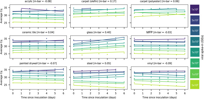
Linear regression of signal intensity (average Cq of viral gene calls) on elapsed time since inoculation (days) for each dilution showed minimal decay of viral RNA on eight of nine surface types over 6 days (Fig. 1). The average decay slope for each surface type (m-bar) did not differ significantly from zero (mean
 =
= 0.0407, standard deviation [SD] =0.210). RT-qPCR signal decayed with time only on glass (m-bar
 =
= 0.401, SD =0.212, differing from the population mean by >1.5 standard deviations).

Scatterplots showing the average Cq values of RT-qPCR viral gene calls for corresponding heat-inactivated viral spike-in over 7 days. Viral spike-in concentrations reported as GEs from ddPCR. Linear regressions of average Cq values on days since inoculation per spike-in were overlaid on the measured data. The average decay slope (m-bar) is reported alongside each surface type.
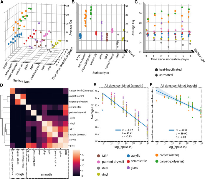
A two-way repeated measure analysis of variance (ANOVA) on viral signal intensity (average Cq) revealed that surface type explains more observed variation in Cq than does time since inoculation at the highest concentration (5 ×
× 105 GEs) (Fig. 2A). A Kruskal-Wallis H test confirmed that mean Cqs differ significantly across surface types (H
 =
= 60.86, P = 2.49
 ×
× 10−9) (Fig. 2B), but not across days since inoculation (H
 =
= 1.34, P = 0.97) (Fig. 2C). Pairwise Mann-Whitney U tests comparing ranked values of Cqs from samples grouped by surface type highlight that both carpet materials (olefin and polyester) are significantly different, after correcting for multiple comparisons (false discovery rate [FDR]-Benjamini/Hochberg, alpha
 =
= 0.005), from all other surfaces, but not from each other (Fig. 2B). Other pairwise, significant differences between materials are summarized in Table S1 in the supplemental material. A clustermap of the U statistic from the pairwise comparisons effectively clusters samples by material properties, with rough surfaces clustering away from smooth ones (Fig. 2D).

(A to C) 3D scatterplots showing distribution of average Cq values of viral gene calls over 7 days for nine different surfaces inoculated with 5 ×
× 105 GEs (nine surfaces for heat-inactivated virus [circles], two surfaces [acrylic and olefin carpet] for infectious virus [diamonds]). The distribution of Cqs differs significantly across surface types (B), but not across days since inoculation (C). (D) Clustermap of the U statistic from pairwise Mann-Whitney U tests between surface types. (E and F) Standard curves relating surface viral load (log2 spike-in) to average Cq values across all time points for smooth (E) and rough (F) surface types.
TABLE S1
Statistically significant differences from pairwise Mann-Whitney U tests between ranked values of average Cqs from viral gene calls grouped by surface type after correction for multiple comparisons (**, FDR-Benjamin/Hochberg, alpha =
= 0.005; N.s. = not significant). Download Table S1, DOCX file, 0.01 MB.
Because RT-qPCR signal intensity for most surfaces was time invariant, time-collapsed linear regression models relating viral spike-in concentration (log2 spike-in) to average Cq act as standard curves for estimating viral load on different monitored surfaces from Cq. After segregating samples based on the qualitative material categories of smooth or rough, linear regressions aggregating all time points yielded one standard curve for smooth surfaces (m = −0.77, b =
= 40.41, r = −0.93) (Fig. 2E) and another for rough surfaces (m = −0.52, b
 =
= 39.90, r = −0.84) (Fig. 2F). The reduced slope of the latter curve stems from higher loss of spiked-in viral signal to the rough surface matrix.
To ensure that viral signal stability was not a consequence of selection for resilient viral particles through heat inactivation, we repeated a subset of experiments using infectious virus (untreated) in a BSL-3 laboratory using the B.1.351/Beta variant of SARS-CoV-2 originally identified in South Africa. Due to space limitations in the BSL-3 facility, the untreated virus experiment only included two surface types, acrylic and carpet (olefin) but used the same dilution series and sampling plan.
Results from untreated and heat-inactivated virus are concordant. Untreated virus samples cluster with respect to surface type rather than virion status (heat inactivated or untreated) (Fig. 2D). When evaluating acrylic and carpet (olefin) samples alone, a Kruskal-Wallis H test shows significant differences in the means of Cqs across all groups when samples are grouped by surface type (H =
= 16.37, P = 0.00095) (see Fig. S1A in the supplemental material), but not when grouped by virion status (H
 =
= 1.96, P = 0.161) (Fig. S1B). Furthermore, linear regression on Cq from paired samples between the heat-inactivated and untreated virus experiments show nearly exact correlation despite the use of different variants (m
 =
= 1.05, r
 =
= 0.97) (Fig. S1C).
FIG S1
(A) Swarm plot showing distribution of average Cqs of viral gene calls for acrylic and carpet (olefin) surfaces for both heat-inactivated and untreated samples. (B) Swarm plot comparing distribution of average Cqs of viral gene calls for heat-inactivated or untreated samples. (C) Linear regression on Cqs from paired samples between heat-inactivated and untreated samples. Download FIG S1, TIF file, 5 MB.
Discussion.
We show that detecting SARS-CoV-2 RNA on indoor surfaces in environments potentially exposed to COVID-19-infected individuals is effective across a variety of surfaces and a range of initial viral loads. Our swabbing and RT-qPCR methods have greater sensitivity from smooth surfaces (such as MFP—commonly found on desktops—or vinyl flooring) than rough surfaces (carpet). The stability of the viral signal across time limits the ability to estimate when the surface was inoculated but demonstrates that signal can be detected a week postexposure. There is a possibility that viral signal could decay over a longer period of time, but because the motivation behind this study was to improve temporal resolution over shorter periods, this was beyond the scope of the present work. To improve temporal resolution, surfaces swabbed for environmental monitoring should be cleaned with soap and water, following CDC recommendations (13), in order to remove viral signals (12). Previous work with comparable methods for SARS-CoV-2 detection from surfaces demonstrated that washing contaminated objects with household dishwashing detergent for ≥1 min removed enough viral RNA traces so that only 20% of the severely contaminated objects had detectable viral RNA. Furthermore, the average viral load of the washed surfaces was reduced by ~2.5 Cqs in comparison to untreated objects (14). Thus, cleaning monitored surfaces with soap and water improves the probability of distinction between persistent or separate exposures in subsequent SARS-CoV-2 detection events.
Although direct inoculation of surfaces with viral particles does not represent interaction with an infected individual in a real-world scenario, we do directly show that untreated and heat-inactivated SARS-CoV-2 particles have similar detectability and stability across surface types. These findings allow the use of heat-inactivated particles in testing and validating environmental monitoring methods and remove the burden of performing such experiments in BSL-3 laboratories.
TEXT S1
Materials and Methods. Description of materials and methods for experimental design and sample processing. Download Text S1, DOCX file, 0.02 MB.
FIG S2
Line plots showing the average Cqs of RT-qPCR viral signals for positive samples (circles) over 7 days with overlaid scatterplots showing Cqs for inconclusive samples (diamonds). Inconclusive samples increase sensitivity of viral detection through surface swabs, seen as increased data points for low viral spike-ins in comparison to positive samples alone. Viral spike-in concentrations reported as GEs from ddPCR. Download FIG S2, TIF file, 6.6 MB.
TABLE S2
Primer and probe sequences used for digital droplet PCR quantification of viral genome equivalents. Download Table S2, DOCX file, 0.01 MB.
TABLE S3
Individual SARS-CoV-2 target gene-positive criteria. Positive results were called on individual SARS-CoV-2 target genes that had a Cq of <37 with a confidence of >0.7. A positive result on the extraction control gene (MS2) was called when it had a Cq of <37 with a confidence of >0.3. Download Table S3, DOCX file, 0.01 MB.
TABLE S4
RT-qPCR test result-positive criteria. A positive status was called for samples that had at least 2/3 positive calls on the SARS-CoV-2 target genes. An inconclusive status was called on samples with only 1/3 positive calls on the SARS-CoV-2 target genes. A SARS-CoV-2 detected result was called on samples where at least one out of three SARS-CoV-2 targets had a positive call; both positive and inconclusive status samples yielded SARS-CoV-2 detected results. A negative status was called on samples that had a positive call on the control gene and no positive calls on the SARS-CoV-2 target genes. An invalid result was called when neither the control gene nor the viral target genes generated a positive call. Download Table S4, DOCX file, 0.01 MB.
ACKNOWLEDGMENTS
We thank our partner schools and citizen scientists at 15 sites across five districts in San Diego county.
This research was supported by NIH grant (K08AI130381) and a Career Award for Medical Scientists from the Burroughs Wellcome Fund to A.F.C., NIH grant (K01MH112436) to R.F.-M., and the County of San Diego Health and Human Services Agency (contract 563236). This work was performed with the support of the Genomics and Sequencing Core at the UC San Diego Center for AIDS Research (P30 AI036214), the VA San Diego Healthcare System, and the Veterans Medical Research Foundation. The following reagent was deposited by the Centers for Disease Control and Prevention and obtained through BEI Resources, NIAID, NIH: SARS-related coronavirus 2, isolate USA-WA1/2020, NR-52281. The following reagent was obtained through BEI Resources, NIAID, NIH: SARS-related coronavirus 2, Isolate hCoV-19/South Africa/KRISP-K005325/2020, NR-54009, contributed by Alex Sigal and Tulio de Oliveira.
REFERENCES
Articles from mSystems are provided here courtesy of American Society for Microbiology (ASM)
Citations & impact
Impact metrics
Citations of article over time
Alternative metrics

Discover the attention surrounding your research
https://www.altmetric.com/details/116155979
Smart citations by scite.ai
Explore citation contexts and check if this article has been
supported or disputed.
https://scite.ai/reports/10.1128/msystems.01136-21
Article citations
Exploring indoor and outdoor dust as a potential tool for detection and monitoring of COVID-19 transmission.
iScience, 27(3):109043, 26 Jan 2024
Cited by: 1 article | PMID: 38375225 | PMCID: PMC10875567
Environmental surface surveillance during a COVID-19 outbreak in a school community: implications for controlling indirect transmission.
Braz J Microbiol, 55(1):441-445, 24 Nov 2023
Cited by: 0 articles | PMID: 37996692
Safer at school early alert: an observational study of wastewater and surface monitoring to detect COVID-19 in elementary schools.
Lancet Reg Health Am, 19:100449, 20 Feb 2023
Cited by: 20 articles | PMID: 36844610 | PMCID: PMC9939935
SARS-CoV-2 surveillance with environmental surface sampling in public areas.
PLoS One, 17(11):e0278061, 23 Nov 2022
Cited by: 2 articles | PMID: 36417446 | PMCID: PMC9683569
SARS-CoV-2 RNA Is Readily Detectable at Least 8 Months after Shedding in an Isolation Facility.
mSphere, 7(6):e0017722, 11 Oct 2022
Cited by: 1 article | PMID: 36218344 | PMCID: PMC9769851
Go to all (9) article citations
Data
Data behind the article
This data has been text mined from the article, or deposited into data resources.
BioStudies: supplemental material and supporting data
Similar Articles
To arrive at the top five similar articles we use a word-weighted algorithm to compare words from the Title and Abstract of each citation.
End-to-End Protocol for the Detection of SARS-CoV-2 from Built Environments.
mSystems, 5(5):e00771-20, 06 Oct 2020
Cited by: 14 articles | PMID: 33024053 | PMCID: PMC7542562
SARS-CoV-2 Distribution in Residential Housing Suggests Contact Deposition and Correlates with Rothia sp.
mSystems, 7(3):e0141121, 16 May 2022
Cited by: 7 articles | PMID: 35575492 | PMCID: PMC9239251
Measures implemented in the school setting to contain the COVID-19 pandemic
Cochrane Database Syst Rev, 1:CD015029, 17 Jan 2022
Cited by: 26 articles | PMID: 35037252 | PMCID: PMC8762709
Review Free full text in Europe PMC
Funding
Funders who supported this work.
Burroughs Wellcome Fund (1)
Grant ID: Career Award for Medical Scientists
Burroughs Wellcome Fund (BWF) (1)
Grant ID: Career Award for Medical Scientists
County of San Diego Health and Human Services Agency (1)
Grant ID: 563236
HHS | National Institutes of Health (2)
Grant ID: K08AI130381
Grant ID: K01MH112436
HHS | National Institutes of Health (NIH) (2)
Grant ID: K08AI130381
Grant ID: K01MH112436
NIAID NIH HHS (2)
Grant ID: K08 AI130381
Grant ID: P30 AI036214
NIGMS NIH HHS (1)
Grant ID: T32 GM127235
NIMH NIH HHS (1)
Grant ID: K01 MH112436

 f
and
f
and 

