Abstract
Free full text

Advances in Analysis of Low Signal-to-Noise Images Link Dynamin and AP2 to the Functions of an Endocytic Checkpoint
Associated Data
SUMMARY
Numerous endocytic accessory proteins (EAPs) mediate assembly and maturation of clathrin-coated pits (CCPs) into cargo-containing vesicles. Analysis of EAP function through bulk measurement of cargo uptake has been hampered due to potential redundancy among EAPs and, as we show here, the plasticity and resilience of clathrin-mediated endocytosis (CME). Instead, EAP function is best studied by uncovering the correlation between variations in EAP association to individual CCPs and the resulting variations in maturation. However, most EAPs bind to CCPs in low numbers, making the measurement of EAP association via fused fluorescent reporters highly susceptible to detection errors. Here, we present a framework for unbiased measurement of EAP recruitment to CCPs and their direct effects on CCP dynamics. We identify dynamin and the EAP-binding α-adaptin appendage domain of the AP2 adaptor as switches in a regulated, multistep maturation process and provide direct evidence for a molecular checkpoint in CME.
INTRODUCTION
Clathrin-mediated endocytosis (CME) is a major pathway for internalizing cell surface proteins and receptors (Ferguson and De Camilli, 2012; McMahon and Boucrot, 2011; Traub, 2009). During this process, clathrin and the adaptor protein AP2 assemble into clathrin-coated pits (CCPs) on the plasma membrane, which invaginate and mature with the help of other endocytic accessory proteins (EAPs) (McMahon and Boucrot, 2011). Upon maturation, the GTPase dynamin catalyzes membrane scission, leading to the formation of cargo-containing vesicles.
Live-cell imaging studies have revealed a remarkable heterogeneity in CCP assembly kinetics within individual mammalian cells (Ehrlich et al., 2004; Loerke et al., 2009; Merrifield et al., 2002; Rappoport et al., 2003; Taylor et al., 2011), with CCP life- time analysis revealing two short-lived “abortive” subpopulations and a longer-lived “productive” subpopulation (Ehrlich et al., 2004; Loerke et al., 2009). Experimental manipulation of cargo concentration (Loerke et al., 2009) and clustering (Liu et al.,2010), as well as small interfering RNA (siRNA) knockdown of a subset of EAPs (Mettlen et al., 2009b), shifted the relative proportions of the three subpopulations, suggesting that transitions between them could be gated by molecular checkpoints monitoring the state of assembly, cargo recruitment, and potentially other physical properties of CCPs, such as curvature. However, whether these checkpoints are the consequence of a purely stochastic process or part of an active control mechanism that monitors CCP maturation remains unknown. In this regard, evidence suggests that dynamin may be involved in regulating CCP maturation in addition to its function in vesicle scission (Loerke et al., 2009; Mettlen et al., 2009a; Sever et al., 2000), although its early role in CME remains controversial (Doyon et al., 2011; Ferguson and De Camilli, 2012; McMahon and Boucrot, 2011).
An extensive network of EAPs has been characterized (Schmid and McMahon, 2007; Taylor et al., 2011), but how these factors contribute to the regulation of CCP maturation and whether they function through a putative checkpoint mechanism is unclear, in part because perturbation of EAPs does not yield an unambiguous phenotype. Indeed, siRNA knockdown studies of individual EAPs generally produce only mild effects on the efficiency of transferrin (Tfn) internalization by CME (Huang et al., 2004). Moreover, despite the fact that many EAPs interact through short peptide motifs with the AP2 α-adaptin appendage domain, siRNA knockdown and reconstitution studies demonstrated that Tfn uptake was fully supported by a truncated α-adaptin lacking this domain (Motley et al., 2006). These studies suggested that redundancy precludes diagnosis of EAP function through bulk measurement of CME efficiency. A potentially more sensitive approach to dissect EAP function is to examine the effect of their association on the dynamics and lifetimes of individual CCPs (Henry et al., 2012; Mettlen et al., 2009b).
CCP lifetimes are measured in time-lapse images of cells expressing fluorescently labeled subunits of clathrin or AP2, using spinning disk confocal or total internal reflection fluorescence microscopy (TIRFM). CCPs, generally smaller than 200 nm in diameter, generate a diffraction-limited fluorescence signal that can be detected and tracked using particle tracking software. Several factors affect the accuracy of the resulting lifetimes: (1) the expression level of the fluorescent reporter, which largely determines the signal-to-noise ratio (SNR); (2) the sensitivity and selectivity of the detection method used to identify CCPs; (3) the capability of the tracking method to interpolate missing information when CCP fluorescence is not detected; (4) the choice of fiducial protein, e.g., clathrin or AP2, and potential perturbation effects of the fluorescent tag; and (5) the susceptibility of the fluorophore to bleaching, which constrains exposure time and excitation intensity.
In the past, labeling has been achieved by overexpressing a fluorescent fusion of clathrin light chain a (CLCa) or of one of the AP2 subunits. Both approaches preserve the stoichiometry of clathrin triskelions and adaptors and have been shown to preserve CME function and dynamics (Ehrlich et al., 2004; Gaidarov et al., 1999; Loerke et al., 2009). However, using genome-edited cells expressing CLCa-red fluorescent protein (RFP) from the endogenous locus, Doyon et al. (2011) reported CCP lifetimes that were approximately half of those measured in cells overexpressing enhanced green fluorescent protein (EGFP)-CLCa. While this result raised the concern that much of the imaging data relying on overexpressed fusion constructs could be invalid, the significantly lower fluorescence signal for endogenously expressed CLCa-RFP versus overexpressed EGFP-CLCa also presents enormous challenges that could prevent accurate acquisition of complete CCP traces.
Here, we report the development and validation of highly sensitive and quantitative imaging approaches to acquire and classify complete CCP trajectories and then use these tools to resolve discrepancies regarding the effects of CLCa overexpression, early dynamin function, and the role of AP2-EAP interactions.
RESULTS
Overexpressed Fluorescent Fusions of Clathrin Light Chain Do Not Inhibit CME
Clathrin is the most abundant CCP protein. Thus, labeled clathrin light chains (CLCs) are optimal markers to follow the dynamics of CME in living cells (Ehrlich et al., 2004; Gaidarov et al., 1999; Loerke et al., 2009). CLCa and CLCb bind interchangeably, yet substoichiometrically (Girard et al., 2005), to clathrin heavy chain (CHC) trimers to form clathrin triskelions. In cell lines overexpressing a fluorescent fusion of CLCa (i.e., EGFP-CLCa), unincorporated CLCs are degraded; thus, endogenous CLCs are downregulated (Figure S1A available online). Hence, the fusion protein effectively outcompetes the endogenous, unlabeled CLCa/b and saturates binding to CHC (Figures S1A–S1C). In contrast, endogenously tagged CLCa-RFP (enCLCa-RFP) is substoichiometrically incorporated (Cocucci et al., 2012), leaving a significant portion of the triskelia without fluorescence and generating much weaker signals for tracking CCPs.
To resolve the discrepancy in CME dynamics observed between cells overexpressing EGFP-CLCa fusions (EGFP-CLCa O/X cells) and genome-edited cells expressing endogenous levels of CLC-RFP (enCLCa-RFP cells), we first replicated earlier control experiments that had shown no measurable effect on Tfn uptake in CLC-overexpressing cells. Contrary to the report of Doyon et al. (2011), we observed no inhibition of Tfn uptake at either low or high levels of adenovirus-mediated overexpression of CLCa or CLCb (Figure S1D), regardless of whether we used N- or C-terminal fusions (Figure S1E) or genes from different species (rat or monkey; Figure S1F). These results confirmed that overexpressed fluorescent CLC fusions do not functionally perturb CME.
Automated Detection of CCPs in Live-Cell Images by TIRFM
One explanation for the reduced lifetimes of CCPs measured in enCLCa-RFP cells is the difficulty in accurately tracking CCP trajectories at lower SNRs. To generate unbiased CCP lifetime distributions at low SNR, we developed a computational analysis framework for the detection of image signals from clathrin-coated structures (CCSs) and compared CCS lifetimes extracted from total internal reflection fluorescence (TIRF) movies of EGFP-CLCa O/X cells with those of cells expressing enCLCa-RFP. Images were acquired at 1 frame × s−1 for 10 min, with illumination conditions chosen to maximize CCS fluorescence with minimal bleaching (see Supplemental Experimental Procedures); the imaging conditions used for enCLCa-RFP cells matched those described in Doyon et al. (2011). Performance analysis on a previously used wavelet-based detection algorithm (Loerke et al., 2009) suggested a significant fraction of false-positive and missed detections, both at the SNR achievable in EGFP-CLCa O/X cells and the lower SNR obtained in enCLCa-RFP cells (Figure 1A). By design, wavelets place relatively weak constraints on the shape and size of the detected objects and the distinction of true- from false-positives requires the use of arbitrary thresholds. In the case of diffraction-limited CCSs, much stronger constraints can be imposed through a model-based detection of signals with the characteristics of a point spread function (PSF). Moreover, model-based approaches allow the computation of uncertainty in the model parameters, which enables selection of valid CCS signals based on unbiased statistical tests. We modeled CCS fluorescence as a two-dimensional Gaussian approximation of the microscope PSF (Cheezum et al., 2001) above a spatially varying local background (see Supplemental Experimental Procedures). The actual detection algorithm encompasses two steps: a filter to identify pixels likely to contain CCS fluorescence and a subsequent model-fitting step performing subpixel localization at each putative CCS position (Figure S2A; Supplemental Experimental Procedures). We used the residuals of the model fit to calculate, for each candidate CCS signal, the uncertainty and statistical significance of the estimated fluorescence amplitude. Among these candidates, signals were selected as valid CCS detections if the amplitude was above a 95th percentile confidence threshold in the local background noise distribution calculated from the residuals (Figures S2B and S2C; Supplemental Experimental Procedures). This approach significantly improves detection sensitivity and selectivity compared to prior approaches developed for particle tracking and super-resolution microscopy (which usually rely on hard thresholds on amplitude-to-background ratio for signal selection; Figure S2D) and resulted in reliable detection of CCS fluorescence in either EGFP-CLCa O/X or enCLCa-RFP cells (Figure 1A; Movies S1 and S2).
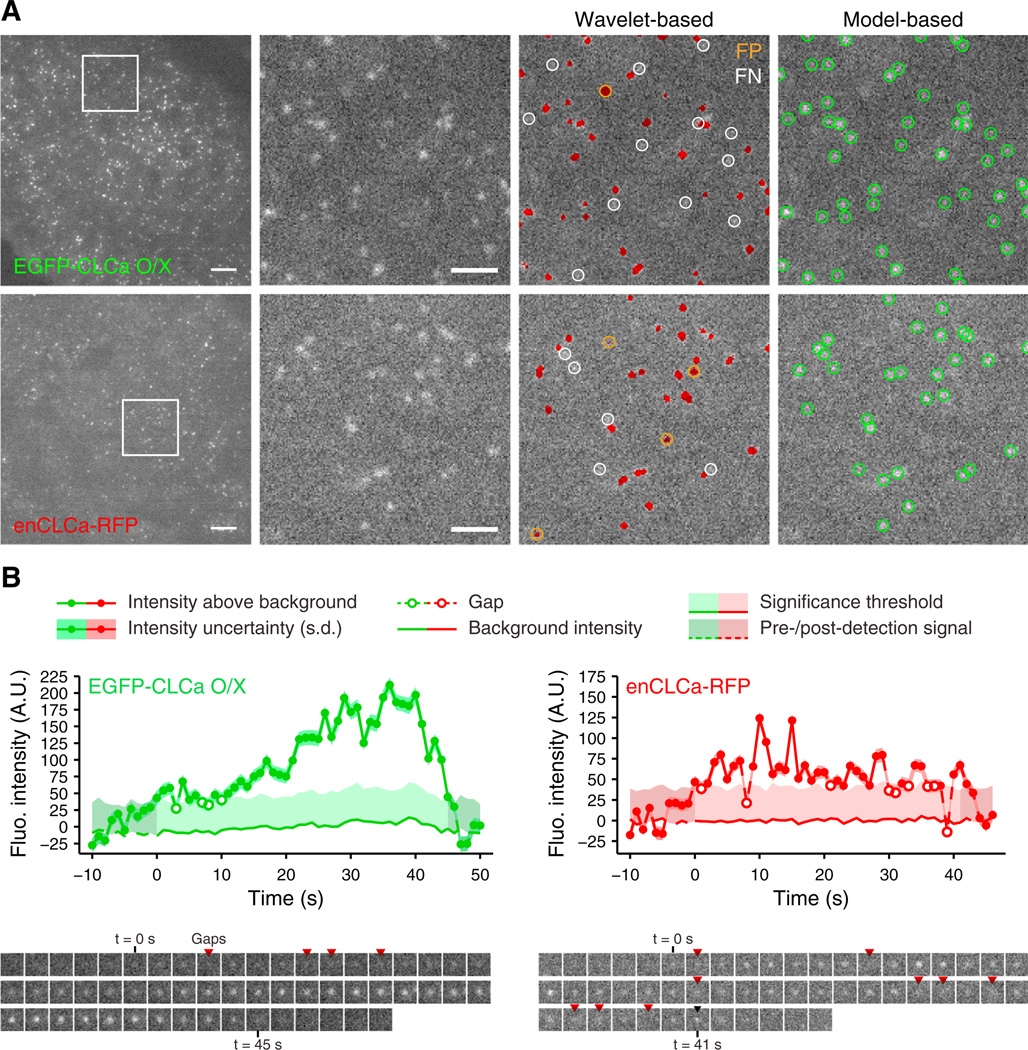
(A) Detection of CCSs in BSC1 cells over-expressing EGFP-CLCa (EGFP-CLCa O/X, top row) or expressing genome-edited, endogenous CLCa-RFP (enCLCa-RFP, bottom row), imaged by TIRFM. Red patches indicate pixels detected as significant by a wavelet-based detection (Loerke et al., 2009); green circles indicate positions of CCSs detected with the model-based algorithm proposed here (see Supplemental Experimental Procedures). False-positives (FP) and false-negatives (FN) of the wavelet-based detection are in reference to the model-based detections. Scale bars: 5 urn, first column, and 2 µm, magnified insets.
(B) Representative intensity traces of CCSs identified by model-based detection and tracking of EGFP-CLCa O/X and enCLCa-RFP. Gaps are defined as frames missed by the detection but recovered during tracking. Uncertainties for the detected intensities (shown as SD, dark shaded band) and the significance threshold (~2 SD above background noise, light shaded area) were estimated using the residuals from the model fit to the raw image signal (see Supplemental Experimental Procedures). Residual signals in the dark shaded regions preceding and following the first and last detected CCS signals, respectively, were measured to verify detection of independent events (see Supplemental Experimental Procedures). Shown below are individual frames of each trace.
See also Figures S1 and S2 and Movies S1 and S2.
To obtain complete fluorescence trajectories of the detected CCSs, we used tracking software to link corresponding locations between consecutive frames as well as to fill in missing detections, or “gaps” (Jaqaman et al., 2008). The latter is critical when the CCS signal fluctuates about the detection limit, as is frequently the case during CCS assembly (Figure 1B). In EGFP-CLCa O/X cells, the traces thus obtained displayed increasing intensity, reflecting CCP growth and maturation (Figure 1B, left panel), whereas in enCLCa-RFP cells, intensity traces were significantly corrupted by noise and often barely persisted above the detection threshold (Figure 1B, right panel).
Robust Measurement of CCP Lifetime Distributions
Equipped with this detection algorithm, we first determined the lifetime distributions of all detected clathrin structures (CSs) in EGFP-CLCa O/X cells (Figure 2A). Relative to previous studies using simpler image processing methods (Ehrlich et al., 2004; Loerke et al., 2009), we detected many more short-lived CSs that emitted very low levels of fluorescence (Figure 2B). Half of the detected events had a lifetime <20 s (Figure 2B, inset). We suspect that these events correspond to the stochastic and transient assembly of small coat fragments. To identify and measure the lifetime distributions of bona fide CCPs, i.e., those undergoing a stabilization and maturation process, we established objective and quantitative criteria to filter out the predominant contribution of these transient CSs.
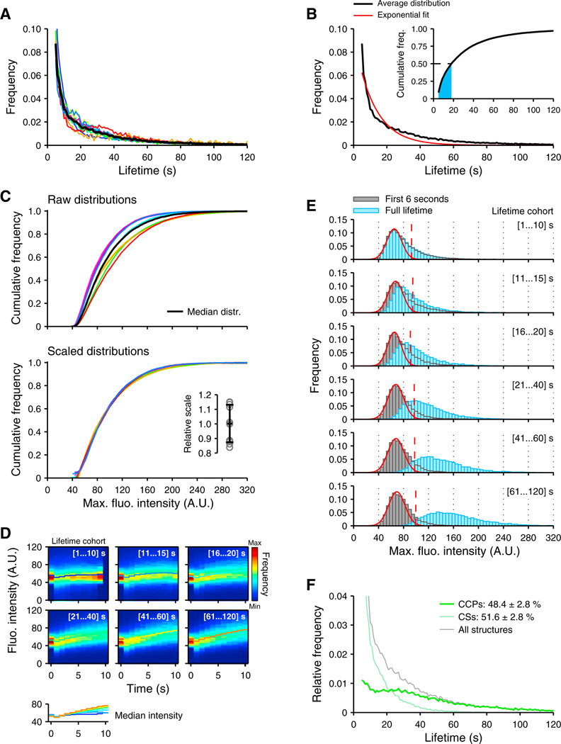
(A) Lifetime distribution of detected CCSs from ten cells overexpressing EGFP-CLCa (colored traces, individual cells; black trace, population average); ~9,000 ± 3,000 CCSs per cell.
(B) Result of the best exponential fit (red) to the average distribution (black; from A). Inset, cumulative distribution. Fifty percent of CCSs have a lifetime <20 s.
(C) Colored traces (top) show cumulative distributions of maximum fluorescence intensity over the lifetime of a CCS in individual cells; black trace, median distribution. After linear scaling (bottom; see Supplemental Experimental Procedures), the distributions narrowly overlay, indicating that the CCS maximum intensity distribution is invariant across cells. Inset, distribution of the scaling factors (mean ± SD, 0.99 ± 0.13).
(D) CCS fluorescence (Fluo.) intensity versus time for different lifetime cohorts. Distributions were calculated from CCSs of ten cells and normalized for each of the ten first time points in the life of a CCS. The median intensity (lower panel) is lifetime invariant during the first ~6 s of assembly. a.u., arbitrary units.
(E) Maximum intensity distributions from scaled CCS intensity traces for different lifetime cohorts. Gray histograms indicate distribution of maximum intensities reached during the first 6 s of a CCS trace. Blue histograms indicate distribution of maximum intensities reached over the full lifetime of a CCS trace. Overlaid red curves indicate best fitting Gaussian distributions to the first mode of the 6 s histograms; dashed lines indicate the 95th percentile of these distributions, which served as a threshold to classify CCSs into transient assemblies (maximum intensity remains below threshold) or bona fide CCPs undergoing maturation (maximum intensity surpasses threshold).
(F) Average lifetime distribution for bona fide CCPs and transient assemblies (CSs) resulting from application of the threshold.
The robust measurement of CCS lifetime distributions generally requires the pooling of measurements from multiple cells (Loerke et al., 2009). Therefore, we first needed to account for variations in the EGFP-CLCa expression level between cells. To this end, we applied an intensity normalization based on the per-cell distributions of the maximum fluorescence emitted by each CCP (Figure 2C, top panel). Scaling each distribution linearly to the median distribution yielded a remarkable overlap between the corrected distributions (Figure 2C, bottom panel; Supplemental Experimental Procedures). The scaling values varied between ~0.9 and 1.1, clearly within the expected variations in EGFP-CLCa expression. Thus, apart from expression level variation, CCP populations follow a stereotypical range of CLCa incorporation.
Next, we grouped CSs into cohorts with a specific range of lifetimes and generated an intensity distribution for each of the first ten detected time points over the CS population in a cohort (Figure 2D). The first detected time point of each CS, regardless of cohort, occurs at the same intensity level (narrow distribution at time 0; Figure 2D), i.e., upon accumulation of the same number of EGFP-CLCa. Subsequently, due to heterogeneity in the CS assembly process, the distributions broaden. Starting at 1 s, they include intensities that are below the detection threshold of the first frame, which were recovered by gap closing during tracking. This representation shows that, regardless of lifetime, the median rate of EGFP-CLCa incorporation during the first ~6 s is identical for all detected structures (Figure 2D). The fluorescence intensity of a majority of CSs in the shortest lifetime cohorts (<10 s and 10–15 s) never exceeded the level reached during this initial phase of assembly (Figure 2E; cf. first 6 s [gray histograms] with total distribution [blue histograms]). In contrast, for longer-lived cohorts, especially those representing CSs with lifetimes >20 s, the intensity distributions beyond 6 s display a systematic increase, indicating the successful nucleation and growth of a nascent CCP. The intensity level of CSs at times <6 s thus provided a threshold for the partitioning of CSs into transient coat fragments versus CCPs that enter a maturation process. To obtain a robust estimate of the threshold, we calculated the Gaussian function best fitting the first peak in the distribution of maximum fluorescence intensities reached in the first 6 s of CS age, across all lifetime cohorts (Figure 2E, red curve; Supplemental Experimental Procedures). The threshold was selected as the 95th percentile of the fitted Gaussian. CSs were then partitioned into two groups: those for which fluorescence intensity exceeds the intensity threshold at any time of the CS trace and those for which fluorescence maximum remains below the threshold. The two populations were of approximately equal size yet had distinct lifetime distributions (Figure 2F). For the population of CSs below the threshold, the distribution showed a rapid, quasi-exponentially decaying process, characteristic of the stochastic disassembly of labile coat structures (Figure S3A), with a mean lifetime of ~14 s. The population of above-threshold, maturing CCPs included all events with a lifetime greater than ~40 s. The maximal intensities of traces in this group varied widely, confirming that the CCP maturation process generates a broad range of coat sizes. Within this population, an anomalous peak of short-lived events (<15 s) remained. We speculated that this population might include events associated with the transient appearance of intracellular membranes coated with clathrin (e.g., originating from endosomes or Golgi, hereinafter termed “visitors”) in the TIRF illumination field. Hence, it was necessary to develop an additional filter that would remove such CME-unrelated signals.
A comparison of the average above-threshold intensities of lifetime cohorts in the CCP population showed that short lifetime cohorts (10–19 s and 20–29 s) included structures that grew at a significantly faster rate than that observed in longer-lived cohorts (Figure 3A). Consistent with the notion that visitors enter the TIRF illumination field as complete assemblies, inspection of the fast-growing events revealed that a majority corresponded to the quasi-instantaneous appearance of a strong signal (Figure 3B). For these events, the intensity in the frames prior to the first detection were at the background level, in contrast to assembling CCPs, which showed a gradual increase in intensity (Figure 3B, blue traces; see also Figure 1B). Exclusion of visitors based on these characteristics eliminated a large fraction of short-lived events from the CCP population. Henceforth, we simply refer to the remaining population of bona fide CCPs as “CCPs.” Their lifetime distribution was characteristic of a process governed by the superposition of multiple steps (Figures 3C and S3B), which may include nucleation, stabilization, maturation, and internalization.

(A) Averaged CCS intensity traces per lifetime cohort, shown as mean ± SE (shaded areas) for all above-threshold CCSs. Inset, enlarged scale showing aberrant rapid rise in intensity of shortlived (<20 s) CCSs (arrowhead).
(B) Examples of intensity traces (green) with instantaneous appearance at an acquisition rate of 1 frame s−1. The majority lasts for less than 10 s, and intensity prior to the first detected frame is at background level, in contrast to CCPs of equally short lifetime (blue).
(C) Subtraction of subthreshold (light green) and transient (light blue) CCSs from all detected CCSs (gray) yields an average lifetime distribution for bona fide CCPs that reflects a multistep maturation process.
See also Figure S3.
Accurate Measurement of CCP Lifetimes Requires High SNR Missed by Endogenous Labels
When applied to EGFP-CLCa O/X and enCLCa-RFP cells, our framework confirmed the previous report (Doyon et al., 2011) in that the lifetimes of CCPs measured with enCLCa-RFP were significantly shorter than those measured with EGFP-CLCa O/X (Figure 4A). Moreover, even after rejection of ~47% of detected CSs as coat fragments, the lifetime distributions in enCLCa-RFP cells still exhibited a quasi-exponential decay. These fundamental differences in CCP lifetime behavior could be related to the difference in CLCa expression levels between the cell types, as suggested by (Doyon et al., 2011), and/or an artificial fragmentation of tracks in enCLCa-RFP cells associated with an increased number of prolonged detection misses due to much lower image SNR (Figure 4A, inset). Indeed, simulations showed that a lifetime distribution corresponding to CCPs in EGFP-CLCa O/X cells could be transformed into a quasi-expo-nentially decaying distribution by as few as one uncompensated gap per track or by gaps occurring with a probability experimentally derived from the analysis of EGFP-CLCa O/X cells (Figure 4B; see Supplemental Experimental Procedures for a discussion of gap-closing accuracy). Thus, we conjectured that much of the apparent CCP lifetime reduction in enCLCa-RFP cells compared to EGFP-CLCa O/X cells is related to detection misses that remained unidentified by our gap-closing algorithm.
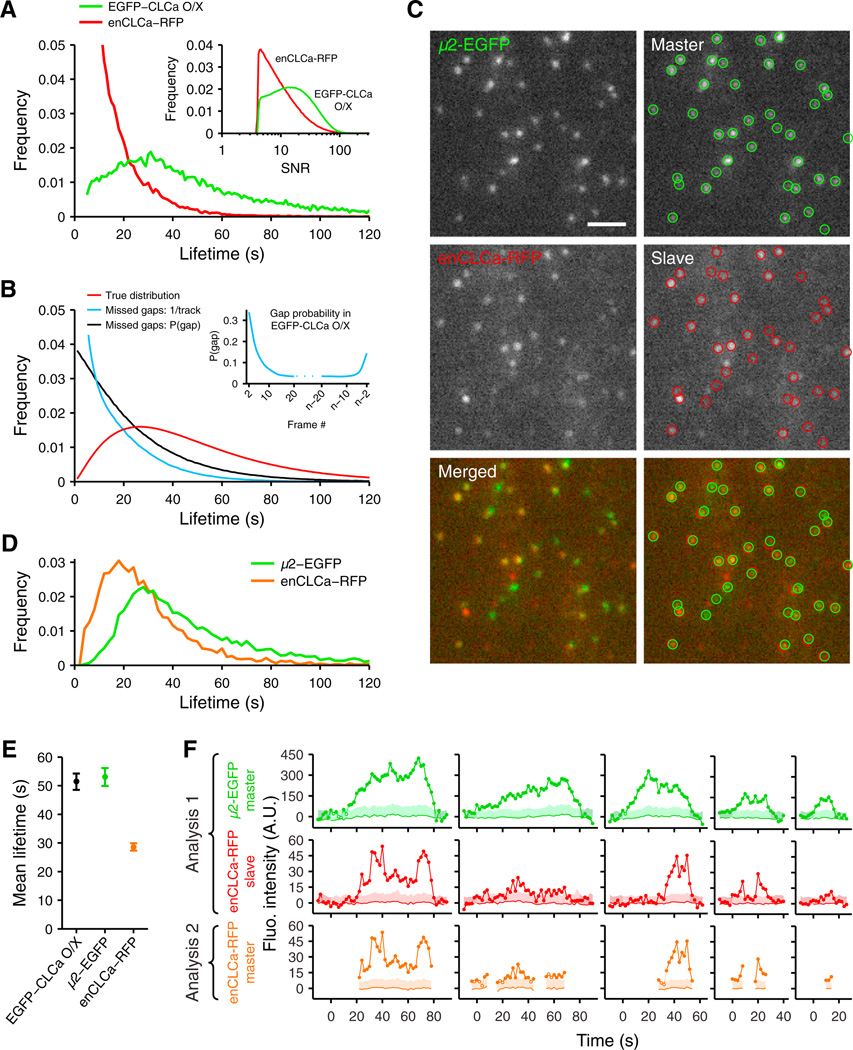
(A) Average lifetime distributions of CCPs for cells overexpressing EGFP-CLCa (green, same data as in Figure 3) and for genome-edited enCLCa-RFP cells (red, average of nine cells). Inset, distribution of SNR for all CCP detections in enCLCa-RFP and EGFP-CLCa O/X cells.
(B) Simulation demonstrating the effects on lifetime distribution of missed detections (gaps) or insufficient gap closing during tracking. Red curve indicates reference gamma distribution fitted to EGFP-CLCa O/X lifetimes in (A) (rate parameter k = 0.05 s−1; shape parameter n = 2.3). Introduction of a single gap with uniform probability, or gap(s) based on the probability of occurrence derived from the EGFP-CLCa O/X data (inset), produces a quasi-exponential lifetime distribution (black and blue lines, respectively, simulated from 106 trajectories).
(C) Master/slave detection of CCSs in genome-edited enCLCa-RFP cells overexpressing µ2-EGFP. Detection was performed on the µ2-EGFP “master” channel (first column); fluorescence intensities in the enCLCa-RFP “slave” channel were estimated by subpixel localization at the detected µ2-EGFP positions (second column; see Supplemental Experimental Procedures). Scale bar, 2 µm.
(D) Lifetime distributions of CCPs measured independently using either the µ2-EGFP or the enCLCa-RFP channel. The enCLCa-RFP distribution was calculated from enCLCa-RFP trajectories with an associated µ2-EGFP signal and vice versa (see Supplemental Experimental Procedures).
(E) Mean CCP lifetimes in EGFP-LCa O/X cells (black) and in enCLCa-RFP cells using the over-expressed µ2-EGFP marker (green) or using enCLCa-RFP (orange). Error bars: cell-to-cell variation, shown as SD from nine cells or more per condition.
(F) Example intensity traces obtained by tracking either µ2-EGFP detections (analysis 1, shown with the associated enCLCa-RFP “slave” signal), or enCLCa-RFP detections (analysis 2).
See also Movie S3.
To test this, we expressed in enCLCa-RFP cells an internal EGFP fusion to the µ2 subunit (µ2-EGFP) of AP2 as a second marker of CCPs (Huang et al., 2003). Similar to CLCa, overexpression of µ2-EGFP outcompetes the endogenous subunit and does not modify the concentration of AP2 or the rate of CME (Ehrlich et al., 2004). We then performed two separate analyses of EGFP/RFP image time-lapse sequences. First, we tracked CCPs in the µ2-EGFP channel (“µ2-EGFP master”) in order to capture complete CCP trajectories, and we measured the associated fluorescence intensity in the enCLCa-RFP channel (“enCLCa-RFP slave”; Figure 4C; Supplemental Experimental Procedures). Next, we tracked CCPs in the enCLCa-RFP channel alone (“enCLCa-RFP master”) and compared the trajectories to those tracked by µ2-EGFP (Movie S3). Due to the transient expression of µ2-EGFP in these experiments, the replacement of endogenous with the tagged version of this subunit was nonsaturating, so that some CCPs had undetectable levels of µ2-EGFP fluorescence. Similarly, due to the low level of enCLCa-RFP relative to unlabeled CLC in these cells (Figure S1C), enCLCa-RFP could not be detected in a fraction of CCPs with detectable µ2-EGFP. To restrict our comparisons to CCP trajectories that were detected in both master channels, we calculated a mutual mapping of the trajectories identified by the two analyses (see Supplemental Experimental Procedures). As hypothesized, the CCP lifetime distribution measured by enCLCa-RFP was skewed toward significantly shorter values, compared with the distribution of the same CCPs measured by µ2-EGFP (Figure 4D). On average, measurements obtained by enCLCa-RFP resulted in an underestimation of lifetimes by a factor of ~1.8 (Figure 4E), which was consistent with the discrepancy reported by Doyon et al. (2011). Moreover, the mean values of the µ2-EGFP and EGFP-CLCa O/X distributions were indistinguishable within the limits of experimental uncertainty, indicating that these two markers are equivalent reporters of CCP dynamics (Figure 4E). Mapping of trajectories from the two analyses allowed us to identify time-shifted fragments that belonged to the same CCP (e.g., second column of panels in Figure 4F). Foremost, this mapping shows that the apparent shorter lifetimes measured by enCLCa-RFP effectively resulted from truncation and/or fragmentation of trajectories, particularly during early time points when the clathrin coat still consists of few triskelia (e.g., Figure 4F, third and fourth columns). Hence, these data demonstrate that the previously reported shorter and more homogeneous lifetimes of CCPs in stable enCLCa-RFP cells versus EGFP-CLCa O/X cells are not the result of perturbations in CCP maturation. Rather, they highlight the critical influence of SNR on lifetime measurements and the need for validation of lifetime distributions by the use of overexpressed, fluorescently tagged CLCa or AP2 subunits as robust fiducial markers of CCPs.
Sensitive Detection of Endogenously Labeled Dynamin-2 Reveals an Early Recruitment and Function for Dynamin in CME
We next applied this analysis to resolve controversies regarding the temporal hierarchy of dynamin recruitment to CCPs. The GTPase dynamin is generally associated with late stages of CME in mammalian cells, where it assembles into collar-like structures around the necks of mature CCPs and catalyzes membrane fission to release clathrin-coated vesicles (CCVs) (Ferguson and De Camilli, 2012; Schmid and Frolov, 2011). Dynamin has also been suggested to function at earlier stages to regulate CCP maturation (Conner and Schmid, 2003; Mettlen et al., 2009a). Indirect evidence for such early roles primarily derives from expression of hypomorphic mutants that block CME at intermediate stages of CCP maturation (Damke et al., 2001), accelerate the rate of CME (Sever et al., 2000), and/or alter the lifetimes of abortive CCPs (Loerke et al., 2009). The observation of dynamin recruitment to nascent CCPs proved more difficult because of the low abundance of labeled dynamin at CCPs, even when expression is driven by a strong ectopic promoter (Soulet et al., 2005; Taylor et al., 2011). Consequently, while some studies report early and/or transient association of dynamin with CCPs throughout their lifetimes (Ehrlich et al., 2004; Mattheyses et al., 2011; Taylor et al., 2012), using endogenously tagged dynamin-2 (enDyn2-EGFP) (Doyon et al., 2011) reported that dynamin recruitment occurred only at very late stages of CCP maturation. Moreover, because 90% of all CCPs exhibited a dynamin recruitment burst, these authors concluded that dynamin must have only minor, if any, functions in regulating CCP maturation. We wondered whether this apparent absence of endogenously labeled dynamin at early stages of CME could also be related to insufficient imaging sensitivity.
To address this, we applied master/slave detection to enDyn2-EGFP cells overexpressing tdTomato-CLCa as the fiducial for CCPs. The tracks obtained from the tdTomato-CLCa master channel were classified as positive for dynamin if the enDyn2-EGFP signal was significant for a duration longer than random association events (see Supplemental Experimental Procedures). As previously reported (Merrifield et al., 2002), dynamin-positive CCPs tend to display a readily detectable accumulation of enDyn2-EGFP ~10–20 s before internalization, likely reflecting the rapid recruitment of dynamin to an assembling constriction ring (Figure 5A). However, when analyzed relative to the tdTomato-CLCa master channel, the readout of the enDyn2-EGFP signal also shows robust association shortly after CCP initiation (Figure 5A; Movie S4). The tdTomato-CLCa master signal enabled the calculation of a significance threshold for enDyn2-EGFP relative to enDyn2-EGFP fluorescence outside of CCPs (Figure 5B; Supplemental Experimental Procedures). Importantly, this threshold shows that, while the early enDyn2-EGFP signal is too weak for independent detection, it remains highly significant when compared against enDyn2-EGFP fluorescence outside of CCPs (Figure 5C; Supplemental Experimental Procedures) or before and after formation of the clathrin coat (Figure 5A, shaded areas).
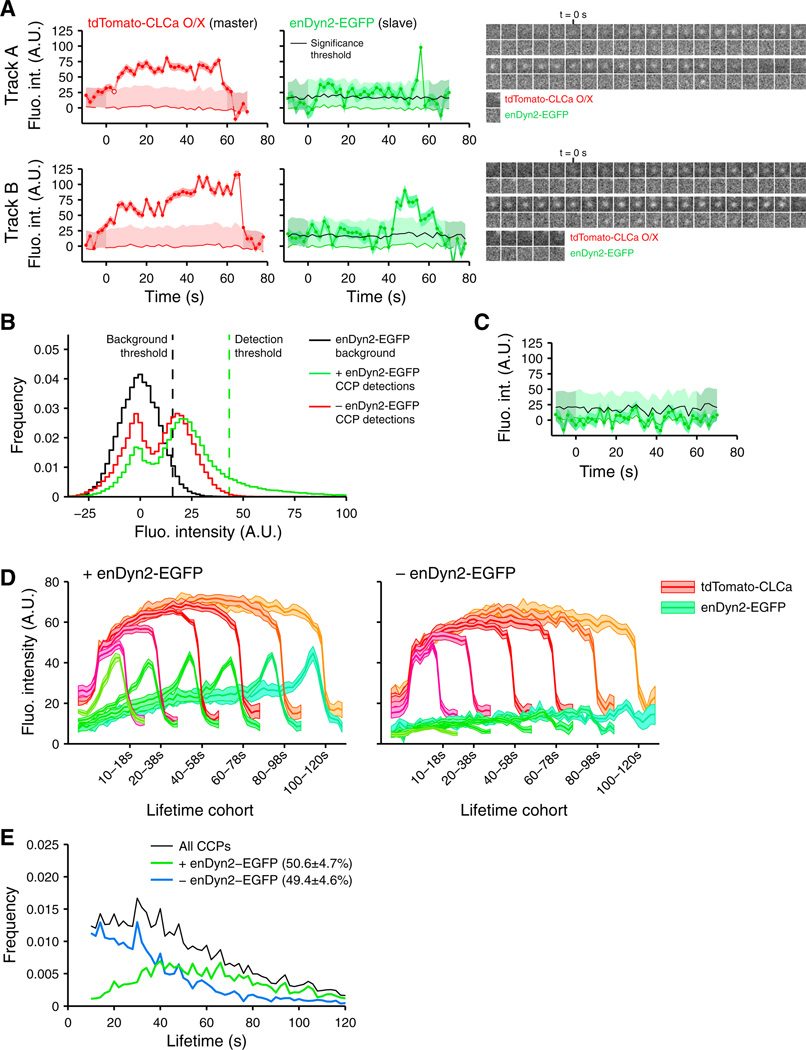
(A) Representative intensity traces of dynamin recruitment early during CCP formation followed by a characteristic peak corresponding to assembly of the scission machinery, measured in SK-MEL-2 cells expressing endogenous Dyn2-EGFP (enDyn2-EGFP) and overexpressing tdTomato-CLCa. Fluo. int., fluorescence intensity.
(B) Distribution of all enDyn2-EGFP slave detections from CCPs with detectable enDyn2-EGFP fluorescence (green) and from CCPs without detectable enDyn2-EGFP (red), compared to the distribution of enDyn2-EGFP outside CCP locations (black; see Supplemental Experimental Procedures). The 95th percentile of the background distribution was selected as a threshold to identify significant enDyn2-EGFP fluorescence [black line in (A)] (see Supplemental Experimental Procedures). A CCP was Dyn2-positive when the number of time points with independently detectable enDyn2-EGFP was significantly above the expected number of such detections due to random association in a trace of equal duration (see Supplemental Experimental Procedures).
(C) Representative trace of enDyn2-EGFP intensity at a random location outside CCSs.
(D) Average clathrin (red tones) and dynamin (green tones) fluorescence intensity traces in lifetime cohorts of Dyn2-positive and Dyn2-negative CCPs (see Supplemental Experimental Procedures). Intensities are shown as mean ± SE calculated from eight cells.
(E) Lifetime distributions of bona fide CCPs identified as described in Figures 2 and and33 and further subcategorized as Dyn2-positive (green) or Dyn2-negative (blue). Characterization of enDyn2-EGFP recruitment relative to CCP disappearance and the full decomposition of lifetimes for CCPs and CSs categorized for enDyn2-EGFP recruitment are shown in Figure S4.
To establish that early recruitment is indeed systematic in all dynamin-positive CCPs, we averaged the enDyn2-EGFP intensity traces for a range of lifetime cohorts (Figure 5D). These averages confirm that dynamin was recruited within 20 s of initiation and accumulates at a steady rate until a rapid burst of recruitment completes the formation of the constriction ring ~12 s before internalization (Figure 5D). Appearance and disappearance-aligned averages of dynamin-positive CCPs show that both the average rate of recruitment during the first ~30 s of maturation and the average timing of the final recruitment burst relative to CCP disappearance were independent of lifetime (Figure S4A). However, at the level of individual CCPs, as previously reported (Mattheyses et al., 2011), we observed significant variation of these parameters, suggesting that the regulation of dynamin recruitment is governed by stochastic processes (Figure 5A).
Unlike the report by Doyon et al. (2011), we also identified a large fraction of dynamin-deficient CCPs (~50%). We compared the lifetime distributions of CCPs with or without dynamin and found significant differences both in their median lifetime (~65 s versus ~30 s) and shape (Figure 5E; for the full decomposition of lifetimes, see Figure S4B). Thus, in the absence of dynamin, the disappearance of CCPs from the plasma membrane is dramatically accelerated. More importantly, the lifetime distribution for dynamin-deficient CCPs displayed a quasi-exponential shape characteristic of a process where the disassembly of structures is triggered by a single random event that occurs at a specific rate (Figure S3A), indicative of abortive events. In sharp contrast, the lifetime distribution of dynamin-positive CCPs peaks at ~40 s and then decays monotonically, indicating that the multistep processes regulating CCP assembly likely act within the first ~40 s of lifetime (Figures S3B and S3C). Together, these analyses establish that dynamin recruitment is a prerequisite for the formation of a productive CCP.
The AP2 α-Adaptin Appendage Domain Regulates Growth and Curvature Induction Early in CCP Assembly
Biochemical, structural, and proteomic studies have shown that the appendage domain of the α-adaptin subunit of AP2 (Figure 6A) is a central interaction hub for a majority of EAPs (Mishra et al., 2004; Motley et al., 2006; Praefcke et al., 2004) and thus should be critical for CCP formation and maturation. Contrary to this expectation, cells expressing a mutant α-adaptin lacking the appendage domain (ΔAD) and siRNA depleted of endogenous α-adaptin had no defect in Tfn uptake (Motley et al., 2006). Thus, the role of AP2-EAP interactions during CCP formation has remained enigmatic, and we wondered whether our analytical approach could help resolve this discrepancy.
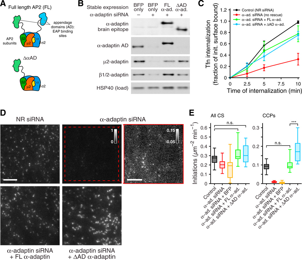
(A) Schematic of the AP2 heterotetramer, showing subunits and appendage domains. The α-adaptin appendage domain (AD) has two characterized binding sites for peptide motifs present in a majority of EAPs (Praefcke et al., 2004).
(B) Immunoblots of AP-2 subunit expression. FL α-adaptin or C-terminally truncated α-adaptin lacking the appendage domain (ΔAD), each harboring siRNA-resistant mutations and bearing a brain-specific insert for detection, were stably expressed in EGFP-CLCa RPE cells. Cells expressing near-endogenous levels of each protein were selected by fluorescence-activated cell sorting, using an internal ribosome entry segment-expressed blue fluorescent protein (BFP). Cells expressing only BFP represent the control condition. The selected cells were subjected to α-adaptin siRNA to silence the endogenous protein.
(C) Tfn uptake for the conditions indicated, shown as mean ± SD of six independent experiments.
(D) EGFP-CLCa-labeled CCSs detected in fixed cells treated as indicated and imaged by TIRFM. All conditions are shown at the same dynamic range, normalized to [0…1], except for the contrast-adjusted α-adaptin siRNA comparison (rightmost panel). Scale bar. 5 µm.
(E) Initiation density of all detected CCSs and bona fide CCPs with lifetime ≥5 s, for the conditions indicated (≥16 cells per condition). Box plots show median, 25th, and 75th percentiles, and outermost data points. ***p < 10−10, permutation test.
To replace α-adaptin at near-endogenous levels, we depleted endogenous α-adaptin by RNA interference in retrovirally transfected EGFP-CLCa retinal pigment epithelial (RPE) cells stably expressing either siRNA-resistant full-length (FL) α-adaptin or the ΔAD α-adaptin (Figure 6B). As previously reported (Motley et al., 2006), Tfn uptake in siRNA-treated cells expressing the ΔAD α-adaptin (ΔAD cells) was comparable to that of untreated control cells or siRNA-treated cells expressing FL α-adaptin (Figure 6C). Knockdown of endogenous α-adaptin in control cells significantly diminished CCP formation, although dim structures could still be detected (Figure 6D, upper right panels). CCP formation was retained in ΔAD cells, but they appeared brighter and more clustered than in control or FL α-adaptin cells (Figure 6D). Using our model-based detection method, we measured the rates of initiation of all detectable CSs, which were similar in all cells, even those depleted of AP2 by siRNA (Figure 6E). This result suggests that many of the low-intensity structures may correspond to AP2-independent clathrin assemblies and provides a direct validation of our filtering framework that distinguishes CCPs from coat fragments. In contrast, after filtering the data to measure only bona fide CCPs, initiations were essentially undetectable in siRNA-treated control cells, as expected, and fully rescued by reconstitution with FL α-adaptin (Figure 6E). Unexpectedly, the rate of initiation of CCPs in ΔAD cells was approximately two times greater than in control or FL α-adaptin cells (Figure 6E).
Although the lifetime distributions for all CSs detected in control, siRNA-treated, and FL α-adaptin or ΔAD cells (Figure 7A) showed no discernable differences, we found striking differences in the lifetime distributions of bona fide CCPs (Figure 7B). In cells lacking α-adaptin, essentially no CCPs can be detected, but reconstitution with FL α-adaptin fully restored the lifetime distributions of CCPs. In contrast, a large subpopulation of CCPs that formed in ΔAD cells was short lived, with a quasi-exponentially decaying lifetime distribution. Hence, the α-adaptin appendage domain has a fundamental effect on switching CCP maturation and internalization from an unregulated random process into a regulated, multistep process (cf. Figures S3A and S3B). Most likely, this happens via recruitment of a network of EAPs that facilitate CCP maturation.
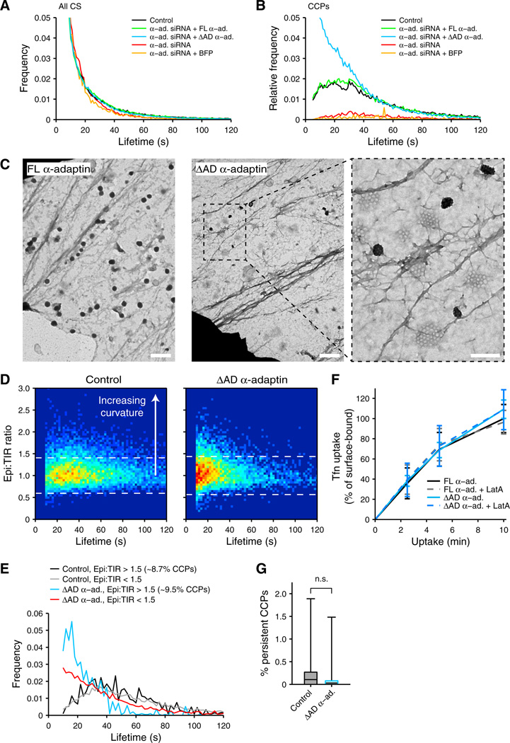
(A) Average lifetime distribution of all detected CCSs in EGFP-CLCa RPE cells for α-adaptin siRNA and re-expression conditions as indicated.
(B) Average lifetime distributions of bona fide CCPs for α-adaptin siRNA and re-expression conditions as indicated.
(C) EMs of “unroofed” RPE cells expressing an siRNA-resistant FL α-adaptin or ΔAD α-adaptin construct after siRNA-mediated depletion of endogenous α-adaptin. Right panel shows higher magnification view of the indicated area. Scale bar, 500 nm; 200 nm for the magnified view.
(D) Epi:TIR ratio intensity levels for individual CCPs plotted as a function of CCP lifetime.
(E) Lifetime distributions of relatively flat (Epi:TIR ratio <1.5) and relatively curved (Epi:TIR ratio >1.5) CCPs in FL α-adaptin-expressing cells (control) and ΔAD α-adaptin-expressing cells.
(F) TfnR internalization in cells expressing FL α-adaptin or ΔAD α-adaptin in the presence and absence of Latrunculin A (100 nM), shown as mean ± SD of four independent experiments.
(G) Percentages of persistent CCPs, which are not included in our lifetime analysis, are not significantly different in FL α-adaptin-expressing cells versus ΔAD α-adaptin-expressing cells. Box plots show median, 25th, and 75th percentiles, and outermost data points. n.s., not significant. p > 0.5, permutation test.
To gain further insight into the role of α-adaptin-EAP interactions in CCP maturation, we acquired electron micrographs (EMs) of the ventral surface of unroofed FL α-adaptin and ΔAD cells (Figure 7C). Although curved coated pits could be detected in both cell populations, ΔAD cells contained a large proportion of small (<200 nm), flat, clathrin lattices rarely detected in FL α-adaptin cells. Many of these appeared in dense clusters spaced below the resolution limit of light microscopy, accounting, in part, for the apparent increase in intensity and clustering seen in fluorescence images (Figure 6D). To test whether these flat structures corresponded to the short-lived CCPs revealed by lifetime measurements, we imaged cells in dual-channel mode, alternating between TIRF and epifluorescence excitation. Because the TIR excitation field decays exponentially, the epifluorescence:TIRF intensity (Epi:TIR) ratio provides a relative measure of coat curvature. We calculated this ratio for each CCP as the maximum detected epifluorescence intensity divided by the maximum detected TIRF intensity, after calibration and adjustment of relative intensities between the two channels (see Supplemental Experimental Procedures). A peak of shortlived (<20 s) flat structures was specifically detected in ΔAD cells, presumably corresponding to the structures seen by EM. We also observed an increased number of curved pits at early time points in ΔAD cells. Hence, CCPs with high curvature seemed to mature significantly faster in ΔAD cells than in wild-type cells (Figure 7D). To examine this more closely, we calculated the 99% confidence interval about the Epi:TIR ratio of 1 expected for a flat structure (Figure 7D, dashed lines) and established 1.5 as a threshold to define highly curved structures (see Supplemental Experimental Procedures). We then calculated lifetime distributions for CCPs with an Epi:TIR ratio >1.5 and compared these to CCPs with an Epi:TIR of <1.5 (Figure 7E). This comparison revealed that, in ΔAD cells, the lifetime distribution of highly curved CCPs displays the characteristics of a multistep process with a very early maturation (peak in lifetime distribution at ~18 s; Figure 7E), while the lifetimes of less curved and flat structures follow a quasi-exponential decay. The median lifetime for curved CCPs (20 s; epi:TIR >1.5) in ΔAD cells was significantly shorter and less variable than for less curved structures (31 s; epi:TIR <1.5). In contrast, no difference was detected between the lifetime distributions of curved versus flatter structures in control cells. These data suggest that the subpopulation of productive CCPs in ΔAD cells undergo a more rapid and less regulated maturation process than those in cells expressing FL α-adaptin.
Paradoxically, ΔAD cells demonstrate a striking phenotype with regard to CCP dynamics (Figure 7B) yet do not exhibit a phenotype with regard to Tfn uptake (Figure 6C). We have identified two compensatory mechanisms in the ΔAD cells that might account, at least in part, for this discrepancy: (1) a 2-fold increase in the rate of CCP initiation that could compensate for the decrease in efficiency of nascent CCP maturation (Figure 6E); and (2) an increase in the rate of maturation of curved CCPs that could account for faster rates of Tfn uptake (Figure 7E), assuming that these highly curved structures corresponded to productive CCPs. However, a third possibility is that Tfn uptake is occurring directly from the flat lattices formed in ΔAD α-adaptin cells. Indeed, previous studies have shown that flat clathrin plaques forming on the adherent surfaces of some cell types are endocytically active and internalized in an actin-dependent manner (Saffarian et al., 2009). However, disruption of actin polymerization with latrunculin A yielded no difference in the rate of Tfn uptake between FL α-adaptin and ΔAD cells (Figure 7F), showing that actin-mediated uptake of flat lattices is not a compensatory mechanism operating in ΔAD cells. We also compared the proportion of persistent structures in ΔAD and control cells and found no statistically significant difference (Figure 7G), indicating that the small flat lattices detected in ΔAD cells are presumably turning over in a nonproductive manner (i.e., that they represent abortive CCPs).
DISCUSSION
We developed a computational framework for sensitive and accurate detection of diffraction-limited objects and described objective criteria for the distinction of bona fide CCPs from transient clathrin assemblies detected in an evanescent field. We applied these tools to three unresolved issues regarding CCP maturation. First, we showed that the apparent lifetime differences of CCPs measured by image data tracking in cells overexpressing EGFP-CLCa versus enCLCa-RFP (Doyon et al., 2011) result only from increased detection errors at the low SNR produced by endogenous labels. Second, we provided direct evidence that dynamin-2 is recruited early during CCP formation and showed that dynamin recruitment is correlated with lifetime kinetics that are characteristic of a regulated maturation process. Third, we showed that the appendage domain of the AP2 α-adaptin subunit, through its multiple interactions with EAPs, plays critical roles at multiple stages in CME, including assembly, curvature generation, stabilization, and maturation of CCPs.
Robust Measurements of CME Dynamics
Model-based identification of diffraction-limited structures combined with refined statistical selection of significant fluorescent signals enabled us to increase the sensitivity of detection of CCPs in live cell images. Applied to endogenous CLCa-RFP and dynamin2-EGFP, we found that, despite the improved performance, the fluorescence levels measurable with endogenously tagged proteins remained too weak to be reliably and consistently detected. Importantly, we show that low signals can be detected with high sensitivity using a master/ slave approach, i.e., by relying on a high-SNR reference such as overexpressed and stoichiometrically incorporated EGFP-CLCa.
EAPs compete for binding to CCP constituents through shared peptide motifs. The EAP-specific affinity for these motifs imparts hierarchy to their recruitment (Mishra et al., 2004), which is therefore particularly sensitive to overexpression. While endogenous labeling avoids such artifacts, a master/slave approach will be critical for the accurate measurement of EAP dynamics at the resulting low SNR. Approaches relying on independent detection of EAP fluorescence are prone to truncation artifacts, especially at the onset of EAP recruitment when the SNR is lowest. Such artifacts are amplified when average recruitment trajectories are calculated from CCPs over the full range of lifetimes, especially after alignment to a single event (Taylor et al., 2011). As our measurements of enDyn2-EGFP recruitment show, recruitment patterns may only become apparent when calculated for defined lifetime cohorts; bulk averages are biased toward the peak recruitment and may obscure lifetime-dependent association patterns.
Dynamin Recruitment during CCP Maturation
Despite evidence for a role of dynamin in the regulation of CCP maturation, most studies have focused on the burst of dynamin recruited at the end of maturation and its function in membrane fission (Ferguson and De Camilli, 2012; McMahon and Boucrot, 2011). Moreover, it has remained unclear to what extent the level of reported early association was correlated to overexpression of fluorescently labeled dynamin (Ehrlich et al., 2004; Taylor et al., 2011, 2012). Using the master/slave approach, we show that endogenous Dyn2-EGFP is consistently recruited within the first 20 s to a subpopulation of CCPs. Independent detection of the dynamin signal would have led to an erroneous conclusion (Doyon et al.,2011).
While the sensitivity of our measurements could not distinguish CCPs that recruited only small amounts of dynamin from those that recruited none, the rapid and presumably abortive turnover of dynamin-deficient CCPs nonetheless suggests that a threshold level of early dynamin recruitment is necessary for CCP maturation. Importantly, subpopulations of dynamin-positive and dynamin-deficient CCPs display not only differences in average lifetime but also a fundamentally different property of lifetime distribution. In the absence of detectable dynamin, the lifetime distribution of CCPs exhibits a quasi-exponential decay reminiscent of a first-order kinetic process where coat disassembly is triggered by a random event occurring at a specific rate (Figure S3A). In the presence of dynamin, the lifetime distribution of CCPs peaks at ~40 s, suggesting that early stages of maturation and CCP survival are actively regulated in a dynamin-dependent manner (Figures S3B and S3C).
Regulation of CCP Maturation by the α-Adaptin Appendage Domain
AP2 complexes act as a prime interaction hub in CME through their ability to bind a majority of EAPs and their function as the link between clathrin and the plasma membrane (Schmid and McMahon, 2007). The finding, reproduced here, that the a-adaptin appendage domain (αAD), which recruits EAPs, was dispensable for efficient CME as measured by Tfn uptake (Motley et al., 2006) was therefore unexpected and has puzzled many researchers in the field. Because many EAPs have the ability to bind to both the ΔAD and β-adaptin appendage domain (βAD), as well as to other CCP constituents, it was assumed that EAP recruitment and CCP maturation were maintained by these redundant interactions. However, our quantitative analysis of CCP dynamics, coupled with EM, has revealed striking pheno-types associated with loss of the αAD.
Contrary to the assumption that clathrin assembly alone drives curvature generation (Dannhauser and Ungewickell, 2012), a significant fraction of nascent CCPs in cells expressing ΔAD α-adaptin fail to gain curvature and form flat, lattice-shaped structures. These aberrant structures are distinct from recently described clathrin plaques in that they are short lived and small (≤200 nm). These data suggest that αAD-EAP interactions may directly or indirectly regulate clathrin assembly into curved lattices. Importantly, curved CCPs that do form in ΔAD cells undergo a more rapid and possibly less complex maturation process than those that form in control cells. Hence, αAD-EAP interactions have a fundamental effect on switching CCP maturation from an unregulated process characterized by first-order, quasi-exponential decay kinetics into a regulated, multistep process (Figure S3). AP2-interacting EAPs could regulate clathrin assembly (e.g., auxilin), generate and/or stabilize curvature (e.g., FCHo1/2, amphiphysin, endophilin, SNX9, epsin), trigger conformational changes in AP2 to enhance cargo binding (e.g., AAK1), interact with cargo molecules (e.g., SNX9, epsin, eps15), and/or act as scaffolds (intersectin, epsl 5). Any of these functions could contribute to the observed requirement for αAD during the early and/or late stages of CME. Determining the identity and hierarchy of EAPs that are essential to these functions will require further studies. Given the robustness of bulk CME to the loss of EAP interactions, the assays described here will be essential for elucidating the specific functions of EAPs.
Plasticity of CME
We have identified two compensatory mechanisms that together are likely to contribute to the unexpected robustness of CME in ΔAD cells. First, the initiation rate for bona fide CCPs was approximately two times higher, potentially compensating for the higher failure rate of abortive structures in these cells. This suggests the existence of some feedback mechanism linking CCP stabilization to initiation rates and/or that αAD somehow negatively regulates the efficiency of CCP nucleation events. Second, although nonproductive, flat lattices constitute a significant proportion of CCSs in ΔAD cells, our EM and Epi:TIR ratio-based analyses reveal a significant fraction of CCPs that appear to assemble normally. CCPs that gain curvature in ΔAD cells appear to undergo maturation much more quickly and synchronously than in control cells, which could also compensate for the loss of αAD-EAP interactions. Importantly, the identification of these compensatory mechanisms and the plasticity of CME have profound implications for future studies of the mechanisms governing CME. One example of the consequence of this plasticity derives from the recently published results of a whole genome siRNA screen based on CME of Tfn: none of the 92 hits identified in this screen corresponded to known EAPs (Kozik et al., 2013). The assays we have developed will be essential for identifying the function of individual EAPs.
CCP Maturation Is Controlled by Regulatory Checkpoints that Depend on Dynamin-2 and Full-Length AP2
The shape of the CCP lifetime distributions revealed by our measurements (Figure 3C) suggests that CCP internalization can be triggered only after completion of one or more rate-limiting, regulatory steps during CCP maturation (Figure S3). The peak of the distribution indicates that key regulating steps likely occur prior to the first ~30–40 s of maturation. Indeed, the distinct lifetime distributions measured for dynamin-positive and dynamin-deficient CCPs support this hypothesis. Dynamin-positive CCPs displayed the characteristics of a multistep process with a majority of lifetimes >40 s. In contrast, the lifetime distribution of dynamin-deficient CCPs decayed quasi-exponentially, consistent with an abortive process undergoing unregulated disassembly with first-order kinetics. Analogous to this observation, the distributions of flat lattices in ΔAD cells are shifted toward shorter lifetimes and display a similar quasi-exponential decay. In contrast, the lifetime distributions of curved pits in ΔAD cells peak at ~20 s, suggesting a more rapid, and potentially less regulated process.
These data are consistent with a checkpoint model that we and others have previously proposed for CME (Henry et al., 2012; Loerke et al., 2009; Puthenveedu and von Zastrow, 2006). Prior evidence for this model includes a statistical decomposition of CCP lifetimes that showed a differential response of abortive versus productive CCP subpopulations to perturbation of dynamin function and cargo concentration (Loerke et al., 2009) and the identification of cargo-specific sub-populations of CCPs with distinct maturation kinetics (Henry et al., 2012; Liu et al., 2010; Puthenveedu and von Zastrow, 2006). There is also evidence that different EAPs (Mettlen et al., 2009b), as well as lipid kinases and phosphatases (Antonescu et al., 2011), may hierarchically act as inputs to the checkpoint. The assays we describe here provide the tools to further test this hypothesis and identify the role of individual EAPs in the regulation of CME.
EXPERIMENTAL PROCEDURES
Cell Biology
The procedures used for culture of human RPE cells, siRNA transfection.CHC immunoprecipitation, and immunoblotting are described in the Supplemental Experimental Procedures available online. Tfn receptor internalization assays were performed as previously described elsewhere (Yarar et al., 2005) and are summarized in the Supplemental Experimental Procedures.
Image Acquisition
TIRF microscopy was performed as previously described elsewhere (Liu et al., 2010). Detailed acquisition parameters are provided in the Supplemental Experimental Procedures.
Image and Data Analysis
The methods for model-based detection of CCSs, validation of CCS trajectories, cell-to-cell intensity normalization, identification of bona fide CCPs, master/slave-based identification of significant EAP signals, calculation of lifetime distributions, and curvature measurement by Epi:TIR ratio are detailed in the Supplemental Experimental Procedures. CCS tracking was performed using the u-track software package (Jaqaman et al., 2008), and all trajectories representing fully observable (i.e., not truncated by the acquisition), diffraction-limited CCSs were retained for further analysis. All analyses were implemented in Matlab (MathWorks, Natick, MA, USA). The software is available for download as Supplemental Software and at http://lccb.hms.harvard.edu/software.html.
ACKNOWLEDGMENTS
This research was supported by National Institutes of Health grants R01 GM73165 (to G.D. and S.L.S.) and MH61345 (to S.L.S.) and fellowships from the Swiss National Science Foundation (to F.A.) and the American Heart Association (to M.M.). We thank M. Wood, Director of The Scripps Research Institute Electron Microscopy Core, for EM sample preparation and imaging, and M. Leonard and V. Lukiyanchuk for technical assitance. SK-MEL-2 (Human Skin Melanoma) cells expressing CLCa-RFP and/or Dyn2-EGFP under their endogenous promoters were kindly provided by D. Drubin (University of California, Berkeley). We thank T. Kirchhausen (Harvard Medical School) for providing the dtTomato-CLCa construct and BSC1 cells stably expressing EGFP-CLCa, and we thank A. Sorkin (University of Pittsburgh Medical School) and M. Robinson (Cambridge Institute for Medical Research) for kindly providing µ2-EGFP and siRNA-resistant α-adaptin constructs.
Footnotes
SUPPLEMENTAL INFORMATION
Supplemental Information includes Supplemental Experimental Procedures, four figures, four movies, and Supplemental Software and can be found with this article online at http://dx.doi.org/10.1016/j.devcel.2013.06.019.
REFERENCES
- Antonescu CN, Aguet F, Danuser G, Schmid SL. Phosphatidylinositol-(4,5)-bisphosphate regulates clathrin-coated pit initiation, stabilization, and size. Mol. Biol. Cell. 2011;22:2588–2600. [Europe PMC free article] [Abstract] [Google Scholar]
- Cheezum MK, Walker WF, Guilford WH. Quantitative comparison of algorithms for tracking single fluorescent particles. Biophys. J. 2001;81:2378–2388. [Europe PMC free article] [Abstract] [Google Scholar]
- Cocucci E, Aguet F, Boulant S, Kirchhausen T. The first five seconds in the life of a clathrin-coated pit. Cell. 2012;150:495–507. [Europe PMC free article] [Abstract] [Google Scholar]
- Conner SD, Schmid SL. Regulated portals of entry into the cell. Nature. 2003;422:37–44. [Abstract] [Google Scholar]
- Damke H, Binns DD, Ueda H, Schmid SL, Baba T. Dynamin GTPase domain mutants block endocytic vesicle formation at morphologically distinct stages. Mol. Biol. Cell. 2001;12:2578–2589. [Europe PMC free article] [Abstract] [Google Scholar]
- Dannhauser PN, Ungewickell EJ. Reconstitution of clathrin-coated bud and vesicle formation with minimal components. Nat. Cell Biol. 2012;14:634–639. [Abstract] [Google Scholar]
- Doyon JB, Zeitler B, Cheng J, Cheng AT, Cherone JM, Santiago Y, Lee AH, Vo TD, Doyon Y, Miller JC, et al. Rapid and efficient clathrin-mediated endocytosis revealed in genome-edited mammalian cells. Nat. Cell Biol. 2011;73:331–337. [Europe PMC free article] [Abstract] [Google Scholar]
- Ehrlich M, Boll W, Van Oijen A, Hariharan R, Chandran K, Nibert ML, Kirchhausen T. Endocytosis by random initiation and stabilization of clathrin-coated pits. Cell. 2004;118:591–605. [Abstract] [Google Scholar]
- Ferguson SM, De Camilli P. Dynamin, a membrane-remodelling GTPase. Nat. Rev. Mol. Cell Biol. 2012;13:75–88. [Europe PMC free article] [Abstract] [Google Scholar]
- Gaidarov I, Santini F, Warren RA, Keen JH. Spatial control of coated-pit dynamics in living cells. Nat. Cell Biol. 1999;1:1–7. [Abstract] [Google Scholar]
- Girard M, Allaire PD, McPherson PS, Blondeau F. Non-stoichiometric relationship between clathrin heavy and light chains revealed by quantitative comparative proteomics of clathrin-coated vesicles from brain and liver. Mol. Cell. Proteomics. 2005;4:1145–1154. [Abstract] [Google Scholar]
- Henry AG, Hislop JN, Grove J, Thorn K, Marsh M, von Zastrow M. Regulation of endocytic clathrin dynamics by cargo ubiquitination. Dev. Cell. 2012;23:519–532. [Europe PMC free article] [Abstract] [Google Scholar]
- Huang F, Jiang X, Sorkin A. Tyrosine phosphorylation of the beta2 subunit of clathrin adaptor complex AP-2 reveals the role of a di-leucine motif in the epidermal growth factor receptor trafficking. J. Biol. Chem. 2003;278:43411–43417. [Abstract] [Google Scholar]
- Huang F, Khvorova A, Marshall W, Sorkin A. Analysis of clathrin-mediated endocytosis of epidermal growth factor receptor by RNA interference. J. Biol. Chem. 2004;279:16657–16661. [Abstract] [Google Scholar]
- Jaqaman K, Loerke D, Mettlen M, Kuwata H, Grinstein S, Schmid SL, Danuser G. Robust single-particle tracking in live-cell time-lapse sequences. Nat. Methods. 2008;5:695–702. [Europe PMC free article] [Abstract] [Google Scholar]
- Kozik P, Hodson NA, Sahlender DA, Simecek N, Soromani C, Wu J, Collinson LM, Robinson MS. A human genome-wide screen for regulators of clathrin-coated vesicle formation reveals an unexpected role for the V-ATPase. Nat. Cell Biol. 2013;15:50–60. [Europe PMC free article] [Abstract] [Google Scholar]
- Liu AP, Aguet F, Danuser G, Schmid SL. Local clustering of transferrin receptors promotes clathrin-coated pit initiation. J. Cell Biol. 2010;191:1381–1393. [Europe PMC free article] [Abstract] [Google Scholar]
- Loerke D, Mettlen M, Yarar D, Jaqaman K, Jaqaman H, Danuser G, Schmid SL. Cargo and dynamin regulate clathrin-coated pit maturation. PLoS Biol. 2009;7:e57. [Abstract] [Google Scholar]
- Mattheyses AL, Atkinson CE, Simon SM. Imaging single endocytic events reveals diversity in clathrin, dynamin and vesicle dynamics. Traffic. 2011;12:1394–1406. [Europe PMC free article] [Abstract] [Google Scholar]
- McMahon HT, Boucrot E. Molecular mechanism and physiological functions of clathrin-mediated endocytosis. Nat. Rev. Mol. Cell Biol. 2011;12:517–533. [Abstract] [Google Scholar]
- Merrifield CJ, Feldman ME, Wan L, Aimers W. Imaging actin and dynamin recruitment during invagination of single clathrin-coated pits. Nat. Cell Biol. 2002;4:691–698. [Abstract] [Google Scholar]
- Mettlen M, Pucadyil T, Ramachandran R, Schmid SL. Dissecting dynamin’s role in clathrin-mediated endocytosis. Biochem. Soc. Trans. 2009a;37:1022–1026. [Europe PMC free article] [Abstract] [Google Scholar]
- Mettlen M, Stoeber M, Loerke D, Antonescu CN, Danuser G, Schmid SL. Endocytic accessory proteins are functionally distinguished by their differential effects on the maturation of clathrin-coated pits. Mol. Biol. Cell. 2009b;20:3251–3260. [Europe PMC free article] [Abstract] [Google Scholar]
- Mishra SK, Hawryluk MJ, Brett TJ, Keyel PA, Dupin AL, Jha A, Heuser JE, Fremont DH, Traub LM. Dual engagement regulation of protein interactions with the AP-2 adaptor α appendage. J. Biol. Chem. 2004;279:46191–46203. [Abstract] [Google Scholar]
- Motley AM, Berg N, Taylor MJ, Sahlender DA, Hirst J, Owen DJ, Robinson MS. Functional analysis of AP-2 α and µ2 subunits. Mol. Biol. Cell. 2006;77:5298–5308. [Europe PMC free article] [Abstract] [Google Scholar]
- Praefcke GJK, Ford MGJ, Schmid EM, Olesen LE, Gallop JL, Peak-Chew SY, Vallis Y, Babu MM, Mills IG, McMahon HT. Evolving nature of the AP2 α-appendage hub during clathrin-coated vesicle endocytosis. EMBO J. 2004;23:4371–383. [Europe PMC free article] [Abstract] [Google Scholar]
- Puthenveedu MA, von Zastrow M. Cargo regulates clathrin-coated pit dynamics. Cell. 2006;127:113–124. [Abstract] [Google Scholar]
- Rappoport JZ, Taha BW, Lemeer S, Benmerah A, Simon SM. The AP-2 complex is excluded from the dynamic population of plasma membrane-associated clathrin. J. Biol. Chem. 2003;278:47357–7360. [Abstract] [Google Scholar]
- Saffarian S, Cocucci E, Kirchhausen T. Distinct dynamics of endocytic clathrin-coated pits and coated plaques. PLoS Biol. 2009;7:e1000191. [Europe PMC free article] [Abstract] [Google Scholar]
- Schmid EM, McMahon HT. Integrating molecular and network biology to decode endocytosis. Nature. 2007;448:883–888. [Abstract] [Google Scholar]
- Schmid SL, Frolov VA. Dynamin: functional design of a membrane fission catalyst. Annu. Rev. Cell Dev. Biol. 2011;27:79–105. [Abstract] [Google Scholar]
- Sever S, Damke H, Schmid SL. Dynamim:GTP controls the formation of constricted coated pits, the rate limiting step in clathrin-mediated endocytosis. J. Cell Biol. 2000;150:1137–1148. [Europe PMC free article] [Abstract] [Google Scholar]
- Soulet F, Yarar D, Leonard M, Schmid SL. SNX9 regulates dynamin assembly and is required for efficient clathrin-mediated endocytosis. Mol. Biol. Cell. 2005;16:2058–2067. [Europe PMC free article] [Abstract] [Google Scholar]
- Taylor MJ, Perrais D, Merrifield CJ. A high precision survey of the molecular dynamics of mammalian clathrin-mediated endocytosis. PLoS Biol. 2011;9:e1000604. [Europe PMC free article] [Abstract] [Google Scholar]
- Taylor MJ, Lampe M, Merrifield CJ. A feedback loop between dynamin and actin recruitment during clathrin-mediated endocytosis. PLoS Biol. 2012;10:e1001302. [Europe PMC free article] [Abstract] [Google Scholar]
- Traub LM. Tickets to ride: selecting cargo for clathrin-regulated internalization. Nat. Rev. Mol. Cell Biol. 2009;10:583–596. [Abstract] [Google Scholar]
- Yarar D, Waterman-Storer CM, Schmid SL. A dynamic actin cytoskeleton functions at multiple stages of clathrin-mediated endocytosis. Mol. Biol. Cell. 2005;16:964–975. [Europe PMC free article] [Abstract] [Google Scholar]
Full text links
Read article at publisher's site: https://doi.org/10.1016/j.devcel.2013.06.019
Read article for free, from open access legal sources, via Unpaywall:
http://www.cell.com/article/S1534580713003821/pdf
Citations & impact
Impact metrics
Citations of article over time
Alternative metrics
Smart citations by scite.ai
Explore citation contexts and check if this article has been
supported or disputed.
https://scite.ai/reports/10.1016/j.devcel.2013.06.019
Article citations
The Nedd4L ubiquitin ligase is activated by FCHO2-generated membrane curvature.
EMBO J, 14 Oct 2024
Cited by: 0 articles | PMID: 39402328
The conserved protein adaptors CALM/AP180 and FCHo1/2 cooperatively recruit Eps15 to promote the initiation of clathrin-mediated endocytosis in yeast.
PLoS Biol, 22(9):e3002833, 24 Sep 2024
Cited by: 0 articles | PMID: 39316607 | PMCID: PMC11451990
Clathrin-associated carriers enable recycling through a kiss-and-run mechanism.
Nat Cell Biol, 26(10):1652-1668, 19 Sep 2024
Cited by: 0 articles | PMID: 39300312
The translesion polymerase Pol Y1 is a constitutive component of the B. subtilis replication machinery.
Nucleic Acids Res, 52(16):9613-9629, 01 Sep 2024
Cited by: 1 article | PMID: 39051562 | PMCID: PMC11381352
Ubiquitin-driven protein condensation stabilizes clathrin-mediated endocytosis.
PNAS Nexus, 3(9):pgae342, 21 Aug 2024
Cited by: 1 article | PMID: 39253396 | PMCID: PMC11382290
Go to all (228) article citations
Data
Data behind the article
This data has been text mined from the article, or deposited into data resources.
BioStudies: supplemental material and supporting data
Similar Articles
To arrive at the top five similar articles we use a word-weighted algorithm to compare words from the Title and Abstract of each citation.
DASC, a sensitive classifier for measuring discrete early stages in clathrin-mediated endocytosis.
Elife, 9:e53686, 30 Apr 2020
Cited by: 16 articles | PMID: 32352376 | PMCID: PMC7192580
Functional characterization of 67 endocytic accessory proteins using multiparametric quantitative analysis of CCP dynamics.
Proc Natl Acad Sci U S A, 117(50):31591-31602, 30 Nov 2020
Cited by: 22 articles | PMID: 33257546 | PMCID: PMC7749282
Regulation of clathrin-mediated endocytosis by hierarchical allosteric activation of AP2.
J Cell Biol, 216(1):167-179, 21 Dec 2016
Cited by: 91 articles | PMID: 28003333 | PMCID: PMC5223608
Regulation of Clathrin-Mediated Endocytosis.
Annu Rev Biochem, 87:871-896, 16 Apr 2018
Cited by: 271 articles | PMID: 29661000 | PMCID: PMC6383209
Review Free full text in Europe PMC
Funding
Funders who supported this work.
NIGMS NIH HHS (2)
Grant ID: R01 GM73165
Grant ID: R01 GM073165
NIMH NIH HHS (3)
Grant ID: MH61345
Grant ID: R37 MH061345
Grant ID: R01 MH061345


